FIGURE

Fig. S4

ID
ZDB-FIG-190604-59
Publication
Lan et al., 2019 - TETs Regulate Proepicardial Cell Migration through Extracellular Matrix Organization during Zebrafish Cardiogenesis
Other Figures
All Figure Page
Back to All Figure Page
Fig. S4

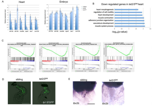
tet2/3DM Larvae Show Cardiac AVC Defects at 48-hpf. Related to Figure 3 and Figure 4.
(A) Quantitative RT-PCR validation of top down-regulated genes from RNA sequencing. RT-PCR analysis ofrdh10a, hoxb8a, rspo2, bax1, cxcl12b transcripts in 2-dpf embryonic heart or whole embryo. Notably, rdh10bapparently compensates for the rdh10a loss, with 4-fold increase in tet2/3DM hearts, which is consistent with RNA sequencing results.
(B) GO analysis shows down-regulated biological pathways in 48 hpf tet2/3DM hearts compared to wildtype hearts by RNA sequencing using isolated hearts.

(C) Gene set enrichment analysis shows down-regulated biological pathways in 48-hpf tet2/3DM hearts compared to wildtype hearts by RNA sequencing using isolated hearts.
(D) Normal Notch Activity in tet2/3DM endocardium. GFP-labeled atrial endocardium indicates Notch activity in sibling hearts as well as tet2/3DM hearts. Hearts were dissected from 48 hpf larvae carrying the Tg(tp1:EGFP) Notch reporter transgene.
(E) WISH for AVC markers tbx2b at 48 hpf. Black arrows indicate AVC specific expression of tbx2b in sibling but not tet2/3DM heart. Scale bars: 50μm.

Expression Data

Expression Detail
Antibody Labeling
Phenotype Data

Phenotype Detail
Acknowledgments
This image is the copyrighted work of the attributed author or publisher, and ZFIN has permission only to display this image to its users. Additional permissions should be obtained from the applicable author or publisher of the image. Full text @ Cell Rep.