
Fig. 5
- ID
- ZDB-FIG-190604-54
- Publication
- Lan et al., 2019 - TETs Regulate Proepicardial Cell Migration through Extracellular Matrix Organization during Zebrafish Cardiogenesis
- Other Figures
- All Figure Page
- Back to All Figure Page
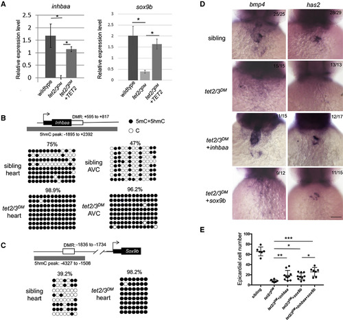
Tet2/3-Dependent Aberrant Promoter Hypermethylation and Deregulation of inhbaa and sox9bLeads to AVC and PE Migration Defects (A) RT-PCR analysis of inhbaa and sox9b transcripts in 48-hpf embryonic heart. (B) DNA methylation status of inhbaa in 48-hpf isolated heartor isolated AVC. Diagram indicates inhbaa locus and the associated regulatory regions. Gray box represents 5hmC peak. Black box represents the coding sequence. White box represents hyper-DMR identified by ERRBS. Profiles of 5mC + 5hmC in hyper-DMR region were validated by bisulfite sequencing. n = 4 per condition. (C) DNA methylation status of sox9b in 48 hpf isolated heart. Diagram indicates sox9b locus and the associated regulatory regions. Gray box represents 5hmC peak. Black box represents the coding sequence. White box represents hyper-DMR. Profiles of 5mC + 5hmC in hyper-DMR region were validated by bisulfite sequencing. n = 4 per condition. (D) WISH for AVC markers bmp4 and has2 at 48-hpf sibling, tet2/3DM, and tet2/3DM injected with inhbaa mRNA or sox9bmRNA larvae. Scale bar: 50 μm. (E) Number of epicardial cells on the heart of 4-dpf sibling, tet2/3DM, and tet2/3DM injected with inhbaa mRNA, sox9bmRNA, or sox9b combined with inhbaa mRNA larvae carrying the Tg(tcf21:NLS-EGFP) transgene. Data are presented as the mean ± SD. The significance is indicated as ∗p < 0.05; ∗∗p < 0.01; ∗∗∗p < 0.001; ns indicates not significant. |
| Genes: | |
|---|---|
| Fish: | |
| Anatomical Terms: | |
| Stage: | Long-pec |
| Fish: | |
|---|---|
| Observed In: | |
| Stage: | Long-pec |