
FIGURE
Fig. 4-S1
Fig. 4-S1
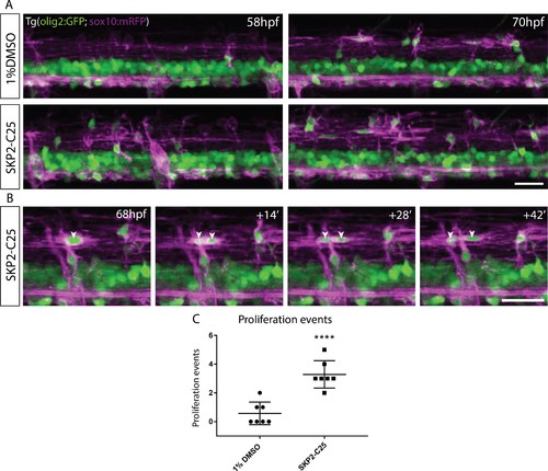
SKP2-C25 increases proliferation of OPCs in the dorsal spinal cord. (A) Frames from time-lapse movies of Tg(Olig2:GFP), Tg(sox10:mRFP) double transgenic control (top) and SKP-C25-treated (bottom) embryos imaged between 58 hpf (left) and 70 hpf (right) Scale bar, 20 μm. (B) Frames from a time-lapse movie of a SKP2-C25-treated animal showing an individual OPC dividing to form two daughter cells (indicated by arrowheads). Scale bar, 20 μm. (C) Analyses of the time-lapse data showed that OPC proliferation events were significantly increased in SKP2-C25 treated animals. |
Expression Data
Expression Detail
Antibody Labeling
Phenotype Data
Phenotype Detail
Acknowledgments
This image is the copyrighted work of the attributed author or publisher, and
ZFIN has permission only to display this image to its users.
Additional permissions should be obtained from the applicable author or publisher of the image.
Full text @ Elife