
Fig. S2
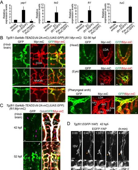
yap1 mRNA expression and Yap1-dependent transcriptional activity in zebrafish endothelial cells (ECs). Related to Figure 2. (A) We isolated EGFP-positive ECs from Tg(fli1:EGFP) embryos and GFP-positive neurons from Tg(huc:GFP) embryos at 2 dpf by fluorescence-activated cell sorting (FACS). Relative mRNA expression of yap1, tie2, fli1, and huc in FACS-sorted cells or whole embryos was analyzed by quantitative PCR (qPCR) analyses. Relative expression is calculated by the expression in the ECs or neurons divided by that in whole embryo. Data from three independent experiments are expressed as mean ± s.d. (n = 3). (B) Projection view of confocal stack fluorescence images of Tg(fli1:Gal4db-TEAD2ΔN-2A-mC);(UAS:GFP);(fli1:Myr-mC) embryos (52-56 hpf). Images of hindbrain and head. Dorsal view, anterior to the top. Images of eye and pharyngeal arch. Lateral view, anterior to the left. GFP images (green), mC images (red), and the merged images are shown. (C) Projection view of confocal images of the hindbrain (lower) in Tg(fli1:Gal4db-TEAD2ΔN-2A-mC);(UAS:GFP);(fli1:Myr-mC) embryos (at 42 and 52 hpf as indicated at the left) injected with Qdot 655 (white) into the heart to visualize perfused vessels. Dorsal view, anterior to the top. Left, GFP images (green); right, the merged images (Qdot 655, white; GFP, green; Myr-mC, red). While a significant population of ECs of perfused CCtA expresses GFP (white arrows), ECs of non-perfused CCtA do not (magenta arrows). (D) Time-sequential two photon stack images during or after lumen formation in ISV of Tg(fli1:EGFP-YAP) embryos (from 42 hpf). Elapsed time is indicated at the top (h:min). Note that EGFP-YAP dynamically shuttles between the nucleus (orange arrowheads) and the cytoplasm (white arrowheads) in lumenized ISV. Similar dynamic behaviors of EGFP-YAP were observed in 3 ISVs of 3 embryos. Scale bars, 10 µm. CCtA, cerebellar central artery; MMCtA, middle mesencephalic central artery; PCS, posterior communicating segment; LDA, lateral dorsal aorta; OV, optic vessel; AA1, mandibular arch; AA2, hyoid arch; AA3, first branchial arch; AA4, second branchial arch; ORA, opercular artery; PHBC, primordial hindbrain channel; BA, basilar artery; ISV, intersomitic vessel. |
Reprinted from Developmental Cell, 40, Nakajima, H., Yamamoto, K., Agarwala, S., Terai, K., Fukui, H., Fukuhara, S., Ando, K., Miyazaki, T., Yokota, Y., Schmelzer, E., Belting, H.G., Affolter, M., Lecaudey, V., Mochizuki, N., Flow-Dependent Endothelial YAP Regulation Contributes to Vessel Maintenance, 523-536.e6, Copyright (2017) with permission from Elsevier. Full text @ Dev. Cell