
Fig. S2
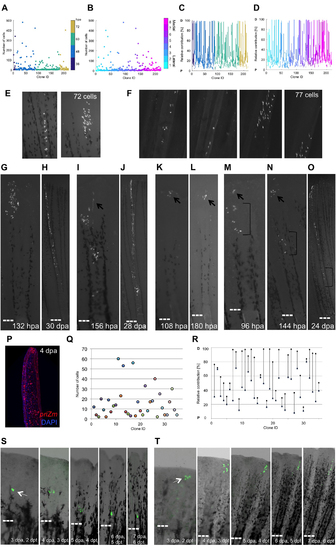
Heterogeneity of Contributions by Blastemal Fibroblast Progenitors, Related to Figure 2 (A) Final number of cells in each clone, sorted by the detection time of initial labeling events: 36 hpa (navy blue), 48 hpa (light blue), 60 hpa (turquoise), or 72 hpa (tan). (B) Final number of cells in each clone, sorted by ray of origin. Cyan (lateral, ray 1) through magenta (medial, ray 8). (C) Relative contribution along the PD axis by individual clones, sorted by the detection time of initial labeling event, as in (A). (D) Relative contribution along the PD axis by individual clones, sorted by the ray of origin, as in (B). P, proximal; D, distal. (E) An example of a clone at 10 dpa represented in 2 images, imaged using a Zeiss AxioZoom microscope. Images were segmented in FIJI software, with a total of 72 cells counted. (F) Zeiss confocal Z-stack images for the same clone as in (E). Images were segmented at each plane in FIJI software, with a total of 77 cells counted. Cell counts from AxioZoom microscope images on average were 95.8% those of counts from optical sections of the same clone (n = 11). (G and H) Clonal expansion through continual proliferation, shown at 132 hpa (G) and 30 dpa (H). Twenty-nine of the tracked clones expanded in this manner. (I and J) Specific morphogenetic process is indicated by preferential creation of one branch of a bifurcated ray, shown at 156 hpa (I) and 28 dpa (J). Arrow in (I) indicates early constituents of the branch. Fifteen tracked clones behaved in this manner. |