FIGURE

Fig. S11

ID
ZDB-FIG-141023-24
Publication
Alexander et al., 2014 - Wnt signaling interacts with bmp and edn1 to regulate dorsal-ventral patterning and growth of the craniofacial skeleton
Other Figures
All Figure Page
Back to All Figure Page
Fig. S11

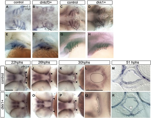
Defects in endoderm and oral ectoderm in dkk1+ embryos (A–L, N–Q) Whole mount ISH, anterior to the left. (A–H) Dorsal and lateral views showing nkx2.3 expression in pharyngeal pouch endoderm at 50 hpf in controls (A, C, E, G), dntcf3+ (B, F), and dkk1+ (D, H) embryos. Normal pouches are numbered. Asterisks and black lines indicate defective pouches. (I–L, N–Q) Ventral views showing pitx2ca expression in control (I–L) and dkk1+ (N–Q) embryos. (M, R) Transverse sections of the mouth showing pitx2ca expression in control (M) and dkk1+ (R) embryos at 51 hpf. Arrowheads indicate expression in oral ectoderm. Dotted lines in K, L, P and Q indicate the mouth opening. White arrowhead indicates a ventral midline fold in the mouth. Scale bars: 100 µm.

Expression Data
Genes:
Fish:
Condition:
Anatomical Terms:
Stage Range: High-pec to Protruding-mouth

Expression Detail
Antibody Labeling
Phenotype Data
Fish:
Condition:
Observed In:
Stage Range: Long-pec to Protruding-mouth

Phenotype Detail
Acknowledgments
This image is the copyrighted work of the attributed author or publisher, and ZFIN has permission only to display this image to its users. Additional permissions should be obtained from the applicable author or publisher of the image. Full text @ PLoS Genet.