
Fig. S4
- ID
- ZDB-FIG-141023-20
- Publication
- Alexander et al., 2014 - Wnt signaling interacts with bmp and edn1 to regulate dorsal-ventral patterning and growth of the craniofacial skeleton
- Other Figures
- All Figure Page
- Back to All Figure Page
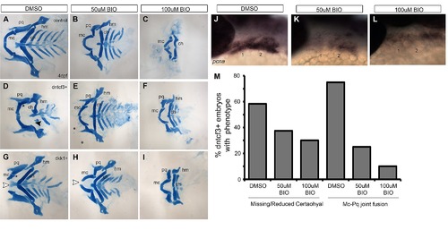
BIO rescues D-V defects in dntcf3+ and dkk1+ embryos. (A–I) Dissected, flat mounted alcian-stained cartilage at 4 dpf, ventral views, anterior to the left; non-transgenic control (top row), dntcf3+ (middle row) and dkk1+ (bottom row), untreated (A, D, G), or treated with 50 µM (B, E, H) or 100 µM (C, F, I) BIO for 6 hours. White arrowheads in G-H indicate Mc clefting, and black arrowheads indicate missing Ch in D. Asterisks in D and G indicate Mc-Pq joint fusions. High concentrations of BIO reduce overall cartilage size and cause specific loss of dorsal cartilages (C). BIO treatments rescue Mc-Pq joint fusions in both dntcf3+ and dkk1+ embryos (E, F, H, I, M) as well as Ch in dntcf3+ embryos (E, F, M). High BIO concentrations rescue Mc clefting in dkk1+ embryos (I). (J–L) Whole mount ISH for pcna in control embryos treated with DMSO, 50 µM BIO, and 100 µM BIO. (M) Histogram quantifying the percentage of dntcf3+ embryos showing Ch loss or Mc-Pq joint fusion in response to BIO treatments. Abbreviations: Ch, ceratohyal; Hm, hyomandibular; Mc, Meckel′s; Pq, palatoquadrate. |