
Fig. 6
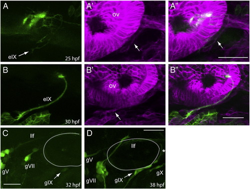
Formation of the IXth nerve is initiated by bmnIX efferents, which are followed by glia and then the gIX afferents. Row A: lateral view of a nkx:mgfp;sox:rfp embryo at 25 hpf (n = 4). The GFP channel (A) shows that the motor efferents from the bmnIX have just entered the periphery at this time, and the arrows in the RFP (A′) and merged (A") channels point to RFP+ peripheral glia that are associating with these axons. Row B: lateral view of a nkx:mgfp;sox:rfp embryo at 30 hpf (n = 5). The GFP channel (B) shows a well-defined efferent fascicle that has just reached the branchial arches, while the RFP (B′) and merged (B") channels show that a definitive sheath (marked by the arrow in B) has encompassed this fascicle. Panel C: lateral view of a 3.2:gfp embryo at 32 hpf shows that the first neurons forming the gIX are just appearing at this time (n = 12). Panel D: lateral view of a 3.2:gfp embryo at 38 hpf shows that the gIX afferents (marked by the asterisk) are just reaching the margin of the hindbrain (n = 11). The dotted line indicates the position of the otic vesicle. gV, trigeminal ganglion sensory neurons; gVII, facial ganglion sensory neurons; gX, vagal ganglion sensory neurons. In all images, anterior is to the left and dorsal at the top. Scale bars are 25 μm. |
| Genes: | |
|---|---|
| Fish: | |
| Anatomical Terms: | |
| Stage Range: | Prim-5 to Prim-25 |
Reprinted from Developmental Biology, 357(2), Cox, J.A., Lamora, A., Johnson, S.L., and Voigt, M.M., Diverse mechanisms for assembly of branchiomeric nerves, 305-17, Copyright (2011) with permission from Elsevier. Full text @ Dev. Biol.