
Fig. 1
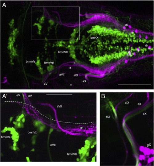
Cranial nerves V, IX and X contain both motor and sensory axons whereas the proximal VIIth is separated into motor or sensory alone. Images shown are confocal z-stacks obtained from 4 dpf larvae in which bmn neurons/axons are green and peripheral sensory axons are magenta. A shows a dorsal view (anterior to left) of a typical 4 dpf isl:gfp;3.2:cherry larva (n = 25). A′ is a higher power image from the same larva (boxed region in A) showing a dorsal view of the motor portion of the VIIth nerve as it exits the hindbrain below the descending trigeminal projections (llf), and of the sensory portion of the VIIth as it enters the hindbrain above the llf. The dashed line marks the approximate position of the hindbrain boundary. B is a lateral view (anterior to left, dorsal at top) caudal to the ear of a typical 4 dpf nkx:mgfp;3.2:cherry larva (nkx:mgfp was used as it contains labeling of bmnIX neurons, unlike the isl:gfp line; n = 7). It shows that the IXth and Xth cranial nerves contain both sensory and motor components at the hindbrain nerve root. This image also shows that the sensory and motor axons are present as separate fascicles within the nerves. bmnVa, anterior motor nucleus of V; bmnVp, posterior motor nucleus of V; bmnVII, motor nucleus of VII; eV, efferent nerve of bmnV; eVII, efferent nerve of bmnVII; eIX, efferent of bmnIX; eX, efferent nerve of bmnX; aV, afferent nerve of gV; aVII, afferent nerve of gVII; aIX, afferent nerve of gIX; aX, afferent nerve of gX; llf, lateral longitudinal fascicle; rb, Rohon–Beard neuron (in magenta); asterisk, peripheral processes of gV and RB neurons. Scale bars; A: 100 μm, A′: 50 μm, B: 20 μm. |
Reprinted from Developmental Biology, 357(2), Cox, J.A., Lamora, A., Johnson, S.L., and Voigt, M.M., Diverse mechanisms for assembly of branchiomeric nerves, 305-17, Copyright (2011) with permission from Elsevier. Full text @ Dev. Biol.