
Fig. 4
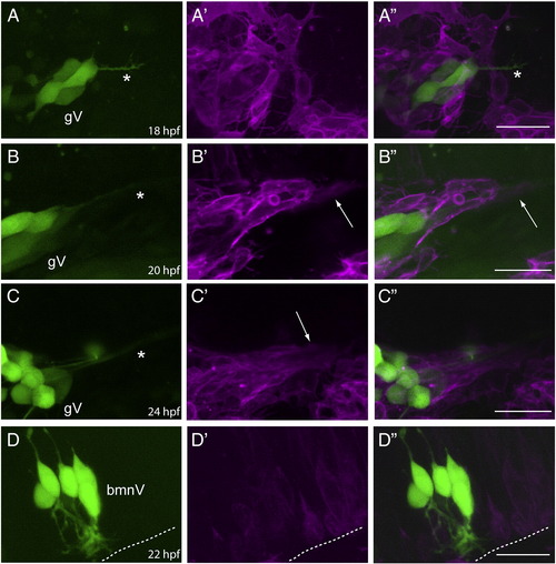
Sensory axons, followed by peripheral glia, are added to the Vth nerve prior to the addition of bmnV efferents. Row A: lateral view of a 3.2:gfp;sox:rfp embryo at 18 hpf (n = 4). The GFP channel (A) shows that the central afferents (marked by the asterisk) from trigeminal ganglion (gV) sensory neurons are growing toward their hindbrain targets, while the RFP (A′) and merged (A") channels show that there are no peripheral glia ensheathing the axon fascicle at this time point. Row B: lateral view of a 3.2:gfp;sox:rfp embryo at 20 hpf (n = 3). The GFP channel (B) shows the central afferents (marked by the asterisk) of the gV sensory neurons as they have extended further toward their hindbrain targets, while the RFP (B′) and merged (B") channels show that the peripheral glia have begun to ensheath the axon fascicle. The arrow in B′ and B" points to the glial sheath that is beginning to form. Row C: lateral view of a 3.2:gfp;sox:rfp embryo at 24 hpf (n = 11). The GFP channel (C) again shows the central afferents (marked by the asterisk) of the gV sensory neurons; the RFP (C′) and merged (C") channels show a well-developed glial sheath surrounding the sensory afferents. The arrow in C′ points to the glial sheath of the Vth nerve. Row D: lateral view of a isl:gfp;sox:rfp embryo at 22 hpf (n = 4). The GFP channel (D) shows that neurons within the Vth motor nucleus are just beginning to extend their efferents; the RFP (D′) and merged (D") channels show that these efferents have not yet extended from the hindbrain into the periphery. The dotted line indicates the margin of the hindbrain. In all images, anterior is to the left and dorsal at the top. Scale bars are 25 μm. |
| Genes: | |
|---|---|
| Fish: | |
| Anatomical Terms: | |
| Stage Range: | 14-19 somites to Prim-5 |
Reprinted from Developmental Biology, 357(2), Cox, J.A., Lamora, A., Johnson, S.L., and Voigt, M.M., Diverse mechanisms for assembly of branchiomeric nerves, 305-17, Copyright (2011) with permission from Elsevier. Full text @ Dev. Biol.