
Fig. 7
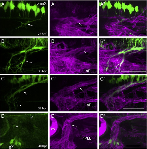
Formation of the Xth nerve is initiated by bmnX efferents, which are followed by glia and then the gX afferents. Row A: lateral view of a isl:gfp;sox:rfp embryo at 27 hpf (n = 5). The GFP channel (A) shows that the vagal motor (bmnX) efferents (marked by the arrow) have entered the periphery, while the RFP (A′) and merged (A") channels show that peripheral glia are associating with the axon fascicles at this time point. Row B: lateral view of a isl:gfp;sox:rfp embryo at 30 hpf (n = 3). The GFP channel (B) shows the bmnX efferents (marked by the arrow) are present as several fascicles, and the RFP (B′) and merged (B") channels show that each set of axons is now enclosed by a defined glial sheath (arrow in B′). Row C: lateral view of a isl:gfp;sox:rfp embryo at 32 hpf (n = 3). The GFP channel (C) shows that the more proximal vagal efferent axons are coalescing to form a distinct fascicle (arrow) whereas the growth cone field of these axons remains diffuse (arrowhead); the RFP (C′) and merged (C") channels show a well-developed glial sheath surrounding the motor efferents. The arrow in C′ points to this Xth nerve peripheral glial sheath. Row D: lateral view of a 3.2:gfp;sox:rfp embryo at 40 hpf (n = 6). The GFP channel (D) shows that gX neurons are just beginning to extend their afferents, with their growth cones (asterisk) having just passed the position of the ganglion of the posterior lateral line; the RFP (D′) and merged (D") channels show that the gX afferents are present within the glial sheath that surrounds the existing bmnX efferents (arrow in D′). In all images, anterior is to the left and dorsal at the top. nPLL, glial sheath of the posterior lateral line nerve. Scale bars are 50 μm. |
| Genes: | |
|---|---|
| Fish: | |
| Anatomical Terms: | |
| Stage Range: | Prim-5 to Prim-25 |
Reprinted from Developmental Biology, 357(2), Cox, J.A., Lamora, A., Johnson, S.L., and Voigt, M.M., Diverse mechanisms for assembly of branchiomeric nerves, 305-17, Copyright (2011) with permission from Elsevier. Full text @ Dev. Biol.