
Fig. 5
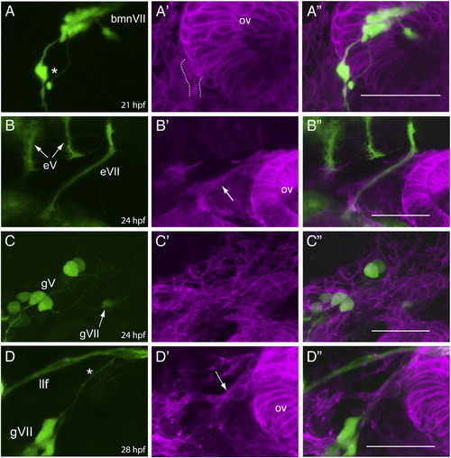
bmnVII axons enter into the periphery and become ensheathed by glia prior to the time when gVII afferents are formed and ensheathed. Row A: lateral view of an isl:gfp;sox:rfp embryo at 21 hpf (n = 6). The GFP channel (A) shows that the VII motor neurons (bmnVII) efferents (marked by the asterisk) are growing toward their peripheral targets, while the RFP (A′) and merged (A") channels show that RFP+ cells are associating with these axons at this time point (dotted lines indicate nerve-associated RFP+ cells). Row B: lateral view of an nkx:mgfp;sox:rfp embryo at 24 hpf (n = 3). The GFP channel (B) shows that the bmnVII axons have grown further in to the periphery, while the RFP (B′) and merged (B") channels show a definitive glial sheath (arrow in B′) has formed around the axon fascicle by this time. Also shown are the processes (marked eV) from the two bmnV nuclei at this stage which are still within the hindbrain, and have not yet joined to form the one efferent bundle that exits the hindbrain at r2. Row C: lateral view of a 3.2:gfp;sox:rfp embryo at 24 hpf (n = 8). The GFP channel (C) shows the gVII sensory neurons (marked by the arrow) are just beginning to form; the RFP (C′) and merged (C") channels show no discernable RFP+ cells are associated with these nascent neurons. Row D: lateral view of a 3.2:gfp;sox:rfp embryo at 28 hpf (n = 5). The GFP channel (D) shows that the afferent VII fibers (marked by the asterisk) have reached the region of the hindbrain margin; the RFP (D′) and merged (D") channels show that these afferents are now ensheathed by peripheral glia (marked by the arrow in D′). gV, trigeminal sensory ganglion neurons; llf, lateral longitudinal fascicle; ov, otic vesicle. In all images, anterior is to the left and dorsal at the top. Scale bars are 50 μm. |
Reprinted from Developmental Biology, 357(2), Cox, J.A., Lamora, A., Johnson, S.L., and Voigt, M.M., Diverse mechanisms for assembly of branchiomeric nerves, 305-17, Copyright (2011) with permission from Elsevier. Full text @ Dev. Biol.