
Fig. 12
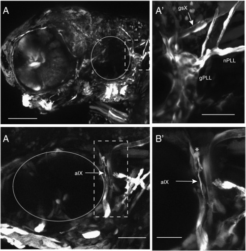
Expression of Foxd3 in cranial sensory neurons does not rescue afferent misrouting in foxd3 null mutants. Panel A, lateral view of 4 dpf mutant foxd3 - / - larva exhibiting a mosaic pattern of Foxd3/GFP co-expressing cells. A′ is higher power image of the boxed region in A and shows that expression of Foxd3 can rescue cranial peripheral glia, as shown by the GFP+ sheaths surrounding the Xth and lateral line nerves (nPLL). In addition, satellite glia are seen in sensory ganglia (gPLL). B: In a different rescued larva, expression of Foxd3 in gIX neurons did not prevent defasciculation of their afferent axons: a higher power image of the boxed region is shown in B′. An asterisk shows misrouted aIX axons that are not sheathed by GFP+ cells. Dotted circle, otic vesicle; gsX, glial sheath of afferent X; gPLL, posterior lateral line ganglion satellite glia; nPLL, posterior lateral line nerve glial cells; aIX, afferent axons of gIX. Anterior to left, dorsal at top. Scale bars: A, 100 μm; A′, 50 μm; B, 50 μm; B′, 25 μm. |
Reprinted from Developmental Biology, 357(2), Cox, J.A., Lamora, A., Johnson, S.L., and Voigt, M.M., Diverse mechanisms for assembly of branchiomeric nerves, 305-17, Copyright (2011) with permission from Elsevier. Full text @ Dev. Biol.