
Fig. S2
- ID
- ZDB-FIG-110407-32
- Publication
- Yee et al., 2011 - Transient receptor potential ion channel Trpm7 regulates exocrine pancreatic epithelial proliferation by Mg2+-sensitive Socs3a signaling in development and cancer
- Other Figures
- All Figure Page
- Back to All Figure Page
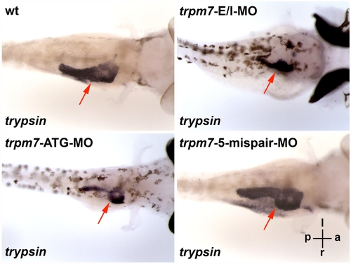
The exocrine pancreas of the swd and trpm7 mutants can be phenocopied by trpm7-E/IMO or trpm7-ATG-MO. The wt embryos injected with non-targeting control MO, trpm7-E/I-MO, trpm7-ATG-MO, or trpm7-5-mispair-MO, were incubated till 96 h.p.f. and analyzed by in situ hybridization using anti-trypsin riboprobes. The wt larvae injected with trpm7-5-mispair-MO appear indistinguishably from those injected with non-targeting control MO, such that for in situ hybridization, they were grown in E3 medium containing PTU that inhibits skin pigmentation, thus facilitating visualization of the exocrine pancreas expressing trypsin. Each larva shown is representative of 10 mutant larvae in each experimental group, and this experiment was repeated with similar results. The orientation of the larvae is indicated: a, anterior; p, posterior; l, left; r, right. The larvae are viewed in the dorsal-ventral direction. The red arrows indicate the trypsin-expressing exocrine pancreas. |