FIGURE

Fig. S1

ID
ZDB-FIG-110407-31
Publication
Yee et al., 2011 - Transient receptor potential ion channel Trpm7 regulates exocrine pancreatic epithelial proliferation by Mg2+-sensitive Socs3a signaling in development and cancer
Other Figures
All Figure Page
Back to All Figure Page
Fig. S1

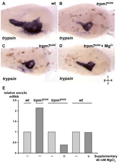
The exocrine pancreas of the trpm7b508 mutant is relatively small, and the defect can be improved by supplementary Mg2+. (A,B) The trpm7b508 mutants and their wt siblings were incubated till 96 h.p.f. and exocrine pancreas analyzed by in situ hybridization using anti-trypsin riboprobes. The wt embryos were grown in medium supplemented with PTU that inhibits skin pigmentation and facilitates visualization of the exocrine pancreas expressing trypsin. (,D) The exocrine pancreas of the trpm7b508 mutants incubated in E3 medium without or with supplementary 40 mM MgCl2 was analyzed on 96 h.p.f. by in situ hybridization using anti-trypsin riboprobes. Each larva shown is representative of 10 mutant larvae in each experimental group, and this experiment was performed two times with similar results. (E) The trpm7b508 mutants and their wt siblings were incubated in E3 medium with or without supplementary 40 mM MgCl2 till 96 h.p.f., and socs3a mRNA was determined by real-time PCR. (C-E) The trpm7b508 mutants were identified based on their hypo-pigmented skin, which was not remarkably affected following incubation with supplementary MgCl2. (A-D) The orientation of the larvae is indicated: a, anterior; p, posterior; l, left; r, right. The larvae are viewed in the dorsal-ventral direction.

Expression Data

Expression Detail
Antibody Labeling
Phenotype Data

Phenotype Detail
Acknowledgments
This image is the copyrighted work of the attributed author or publisher, and ZFIN has permission only to display this image to its users. Additional permissions should be obtained from the applicable author or publisher of the image. Full text @ Dis. Model. Mech.