
Fig. 7
- ID
- ZDB-FIG-100730-9
- Publication
- Eberhart et al., 2006 - Early Hedgehog signaling from neural to oral epithelium organizes anterior craniofacial development
- Other Figures
- All Figure Page
- Back to All Figure Page
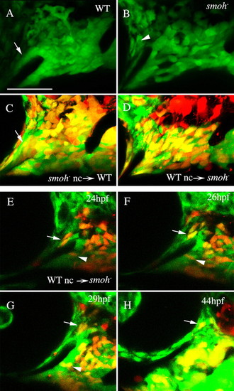
Reception of Hh signaling is not required in neural crest cells to condense on the stomodeum. (A) A tight neural crest cell condensation coats the stomodeum in wild-type 30 hpf fli1:GFP embryos (arrow). (B) The region of this condensation superior to the stomodeum is absent in smo-;fli1:GFP embryos, although GFP-positive sclera is present surrounding the eye (arrowhead). (C,D) 30 hpf embryos following transplantation between wild-type and smo- embryos. (C) smo- neural crest cells readily condense on the stomodeal roof in wild-type embryos (arrow, n=12). (D) Crest cells from wild-type donors fail to condense on the stomodeal roof in smo- embryos, even though they can populate the region occupied by palatoquadrate and Meckel′s cartilage precursors (n=18). (E-H) Images taken from a time-lapse recording (n=2) of wild-type crest in a smo- host. Wild-type neural crest cells are capable of initially populating the region superior to the stomodeum (E-H, arrow); however, these cells are not stabilized in this position and eventually migrate posterior to the eye. Wild-type cells in the Meckel′s cartilage domain condense normally (E-G, arrowhead). Lateral views, dorsal is upwards. nc, neural crest; WT, wild type. Scale bar: 50 μm. |