
Fig. 10
- ID
- ZDB-FIG-100730-12
- Publication
- Eberhart et al., 2006 - Early Hedgehog signaling from neural to oral epithelium organizes anterior craniofacial development
- Other Figures
- All Figure Page
- Back to All Figure Page
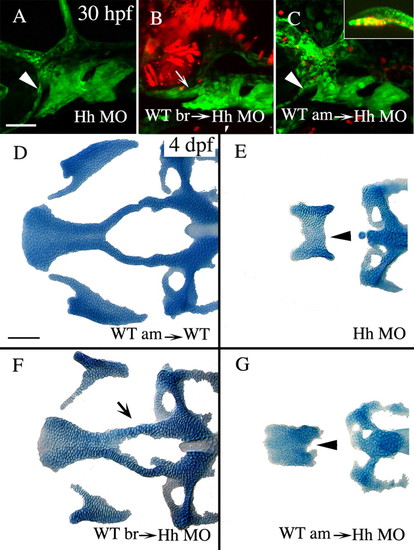
Hh secreted from the ventral presumptive brain, and not the axial mesoderm, is required for condensation of crest cells on the roof of the stomodeum and the subsequent formation of the anterior craniofacial skeleton. (A-C) Lateral views of 30 hpf fli1:GFP embryos co-injected with shh and twhh morpholinos (Hh MO). (D-G) Dorsal views of Alcian stained 4 dpf neurocrania and palatoquadrates from control transplanted embryos (D) or embryos co-injected with Hh MO (E-G). (A,E) Co-injection of Hh MO causes failure of crest cells to condense on the stomodeal roof (A, arrowhead) and the development of the derivatives from this region, the anterior neurocranium and pterygoid process (E, arrowhead; skeletal index=0.1, n=57). (B,F) Transplantation of wild-type brain into Hh MO-injected embryos is capable of rescuing the condensation of neural crest cells (B, arrow) and the skeletal derivatives almost to wild-type morphology (F, arrow, skeletal index=2.2, n=40, ANOVA F ratio=64.481, P<0.0001, Tukey-Kramer shows significance is only attributable to wild-type brain transplant). (C,G) Transplantation of axial mesoderm, including prechordal plate (C, inset), into Hh MO injected embryos has no effect on crest cell condensation (C, arrowhead) or on anterior neurocranium and upper jaw cartilage morphology (G, arrowhead, skeletal index=0.2, n=14). Anterior is leftwards. am, axial mesoderm; br, brain; WT, wild type. Scale bar: 50 μm. |