
Fig. 8
- ID
- ZDB-FIG-100730-10
- Publication
- Eberhart et al., 2006 - Early Hedgehog signaling from neural to oral epithelium organizes anterior craniofacial development
- Other Figures
- All Figure Page
- Back to All Figure Page
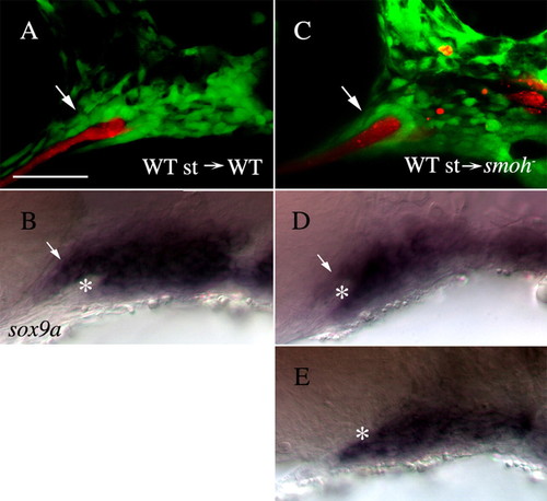
Wild-type stomodeum rescues the condensation of anterior craniofacial neural crest cells in smo- embryos. (A,C) Lateral views of fli1:GFP (A) or smo-;fli1:GFP (C) embryos at 30 hpf following transplantation of wild-type stomodeum. (A) Neural crest cells condense on the stomodeal roof following control transplants (arrow). (C) Following transplantation of wild-type stomodeum into smo-, crest cells condense on the roof of the stomodeum, in a manner similar to that seen in wild type (arrow, n=9). (B,D,E) Lateral views of the same embryos in A,C labeled with RNA probe to sox9a. (B) sox9a-positive cells (arrow) are clearly visible above the stomodeum (*) in control transplanted embryos. (D) sox9a-expressing neural crest cells (arrow) are readily apparent superior to the stomodeum (*) on the side of the embryo receiving the stomodeal transplant. (E) Control, non-transplanted, side of the same embryo as B (image flipped to be the same orientation as that in D). No sox9a-positive cells are observed above the stomodeum (*). Anterior is leftwards. st, stomodeum; WT, wild type. Scale bar: 50 μm. |