
Fig. 5
- ID
- ZDB-FIG-100730-7
- Publication
- Eberhart et al., 2006 - Early Hedgehog signaling from neural to oral epithelium organizes anterior craniofacial development
- Other Figures
- All Figure Page
- Back to All Figure Page
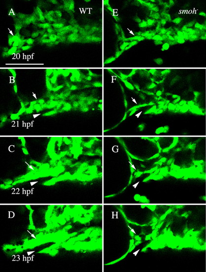
Neural crest cells fail to condense upon the stomodeal roof in smo- embryos. (A-D) Wild-type fli1:GFP embryo. Movie frames are shown every hour from 20 to 23 hpf. Wild-type crest cells are condensing on the stomodeum by 22 hpf (C, arrow) and by 23 hpf (D, arrow) a solid mass surrounding the stomodeum (arrowhead) has formed. (E-H) Neural crest cell behavior in smo- embryos. Crest cells are present just posterior to the eye in smo-;fli1:GFP at 20 hpf (E, arrow). However, these cells fail to condense on the roof of the stomodeum (F-H, arrow). Lateral views, dorsal is upwards. WT, wild type. Scale bar: 50 μm. |