
Fig. 6
- ID
- ZDB-FIG-100730-8
- Publication
- Eberhart et al., 2006 - Early Hedgehog signaling from neural to oral epithelium organizes anterior craniofacial development
- Other Figures
- All Figure Page
- Back to All Figure Page
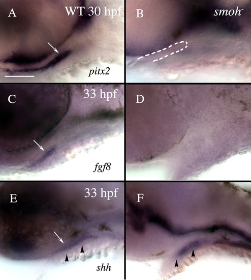
Loss of Hh function causes loss of stomodeum specification. Lateral views of 30 hpf (A,B) or 33 hpf (C-F) wild-type (A,C,E) or smo- (B,D,F) embryos labeled with RNA probe to pitx2 (A,B), fgf8 (C,D) or shh (E,F). (A) Wild-type embryos strongly express pitx2 throughout the stomodeum (arrow), while (B) smo- embryos have no detectable pitx2 expression in the stomodeum, although a morphologically identifiable stomodeum is present (outlined in B). (C) fgf8 labels the lateral stomodeum in wild-type embryos (arrow). (D) No fgf8 expression is detectable in the stomodeum of smo- embryos. (E) shh labels wild-type medial stomodeum (arrow). (F) However, shh is not evident in the stomodeum of smo- embryos, although some medial cells, presumably endoderm, maintain shh expression. Arrowheads in E and F indicate the level of sections shown in Fig. S3. Dorsal is upwards. WT, wild type. Scale bar: 50 μm. |