
Fig. 8
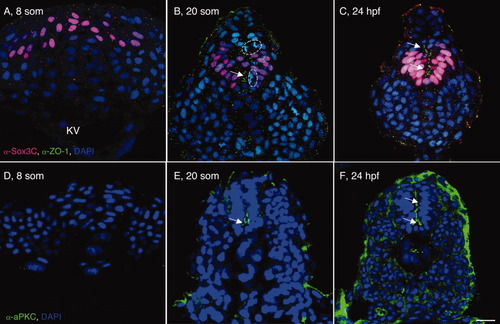
Posterior neural cells undergo progressive epithelialization. A-F: Transverse sections through the tailbud of wild-type (WT) 8 somites (som; A,D), 20 som (B,E), and 24 hours postfertilization (hpf; C,F) embryos labeled with α-Sox3C (A-C, neural tissue, pink), α-ZO-1 (A-C, apical membrane, green), α-aPKC (D-F, apical membrane, green) and DAPI (4′,6-diamidine-2-phenylidole-dihydrochloride; nuclear marker, blue). Arrows point to apical ZO-1 and aPKC localization. Dotted circles surround cells dividing along the mediolateral axis. KV, Kupffer′s vesicle; som, somite; hpf, hr postfertilization. Scale bar = 20 μm. |