
Fig. 7
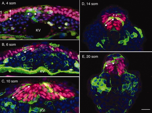
Angular orientation and length-to-width ratios (LWRs) of posterior neural tube (PNT) cells. A-E: Transverse sections through the tailbud of wild-type (WT) embryos at 4 somites (som; A), 6 som (B), 10 som, (C), 14 som (D), and 20 som (E), labeled with membrane-targeted enhanced GFP (mGFP; cell surface marker, green), α-Sox3C (neural tissue, pink) and DAPI (4′,6-diamidine-2-phenylidole-dihydrochloride; nuclear marker, blue). Double arrowheads indicate the angular orientation and length of cells. Asterisks in (D) show cells that are likely to derive from a common mother cell. KV, Kupffer′s vesicle. Scale bar = 20 μm. |