
FIGURE
Fig. 3
Fig. 3
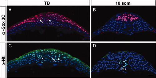
The tailbud is organized in tissue-restricted domains. A-D: Transverse sections of wild-type (WT) tailbud (TB) (A,C) and 10 somites (som; B,D) embryos labeled with α-Sox3C (neural tissue, pink; A,B), α-Ntl (axial mesoderm precursors, green; C,D) and DAPI (4′,6-diamidine-2-phenylidole-dihydrochloride; nuclear marker, blue). Arrows point to clusters of cells that express either low levels of Sox3C (A) or that are Ntl-positive (B). NC, notochord. Scale bar = 20 μm. |
Expression Data
Expression Detail
Antibody Labeling
Phenotype Data
Phenotype Detail
Acknowledgments
This image is the copyrighted work of the attributed author or publisher, and
ZFIN has permission only to display this image to its users.
Additional permissions should be obtained from the applicable author or publisher of the image.
Full text @ Dev. Dyn.