
Fig. 6
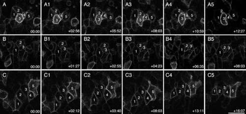
Cell behaviors driving the early stages of neural convergence in posterior regions. Still frames from a time-lapse movie of a wild-type (WT) embryo, viewed from the dorsal side and labeled with membrane-targeted enhanced GFP (mGFP). Anterior is to the right in all panels. A-C: The midline is toward the bottom in series A and B; and toward the top in series C. Panel series A, B, and C illustrate clusters of cells whose behaviors were tracked over time. The embryo was approximately 4 somites (som) at time 0. Time elapsed (hr:min) from the beginning of the movie is indicated in the lower right corner. Scale bar = 20 μm. |