
Fig. 2
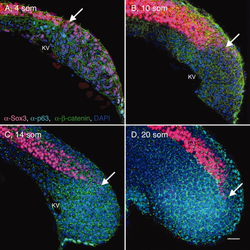
Elongation of the posterior neural tube (PNT). A-D: Sagittal sections through the tailbud (at the level of KV) of wild-type (WT) 4 som (A), 10 som (B), 14 som (C), and 20 som (D) embryos, labeled with α-Sox3C (neural tissue, pink), α-p63 (epidermis, nuclear labeling, turquoise), α-β-catenin (cell membrane labeling, green), and DAPI (4′,6-diamidine-2-phenylidole-dihydrochloride, nucleus, blue). Anterior is to the left and dorsal side is up in these and all subsequent images of sagittal sections. White arrows indicate the caudal-most extent of the Sox3C high-expression domain. KV, Kupffer′s vesicle; som, somites. Scale bar = 20 μm. |