|
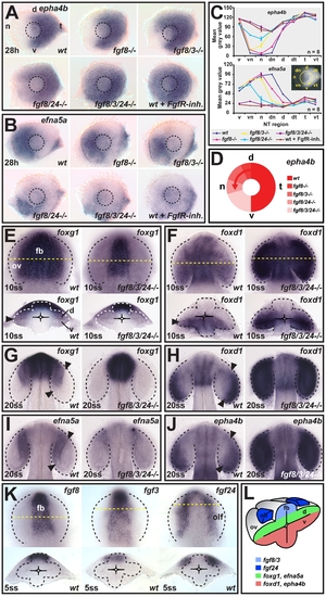
fgf8/3/24 impose nasal-temporal pattern by signaling along the dorsal-ventral axis of the optic vesicle. (A and B) Expression of (A) epha4b in the temporal and (B) efna5a in the nasal retina of wild-type (wt) control, fgf8 mutant (fgf8-/-), fgf8/3 double mutant (fgf8/3-/-), fgf8/24 double mutant (fgf8/24-/-), fgf24-morpholino injected fgf8/3 double mutant (fgf8/3/24-/-), and wt embryos after Fgf receptor inhibitor treatment (wt+FgfR-inh.) at 28 h (nasal is to the left and dorsal is up). Dashed-line circle indicates position of the lens. (C) Nasal-temporal profile of epha4b and efna5a gene expression levels (mean grey value for n = 8 images analyzed for each genotype, y-axis, in defined nasal-temporal (NT) regions of the retina, x-axis, see inset, one representative standard deviation is plotted). (D) Schematic illustrating the graded, nasal expansion of the temporal marker epha4b in the phenotypic series of fgf mutants. (E and F) Expression of the future nasal marker foxg1 (E) and the future temporal marker foxd1 (F) at 10ss in the optic vesicle (ov) of wt (left) and fgf8/3/24-/- embryos (right) (top: dorsal view, anterior to the top, bottom: cross-section, dorsal to the top). (G–J) Expression of the future nasal markers foxg1 (G) and efna5a (I) and the future temporal markers foxd1 (H) and epha4b (J) in the optic cup of wt (left) and fgf8/3/24-/- (right) embryos at 20ss (dorsal views, anterior to the top). Asterisk in (I): remnant efna5a expression in the optic stalk region. (K) Expression of fgf8 and fgf3 in the dorsal forebrain (fb), and fgf24 in the olfactory placode (olf) in wt embryos at 5ss. Orientation as in (E). (L) Schematic cross-section, illustrating dorsal expression of fgfs and expression of future nasal-temporal retina markers along the dorsal-ventral optic vesicle axis at 10ss. Arrowheads in (E–J): gene expression limits in the optic vesicle and cup. Yellow dotted lines indicate transverse section level. Black and white dotted lines indicate neural tube or optic vesicle/cup outlines. d, dorsal; dn, dorsonasal; dt, dorsotemporal; n, nasal; t, temporal; v, ventral; vn, ventronasal; vt, ventrotemporal.
|