
Fig. 3
- ID
- ZDB-FIG-091214-19
- Publication
- Picker et al., 2009 - Dynamic coupling of pattern formation and morphogenesis in the developing vertebrate retina
- Other Figures
- All Figure Page
- Back to All Figure Page
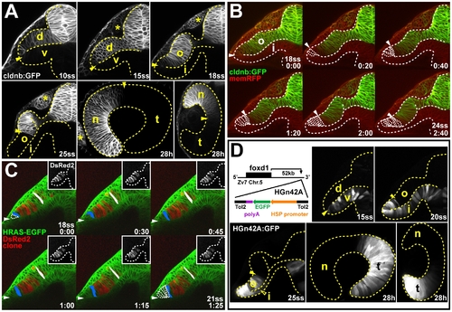
In vivo imaging reveals late movement of prospective temporal retinal cells around the distal ridge of the optic cup. (A) GFP expression (white) in the optic vesicle and optic cup of live Tg(-8.0cldnb:lynGFP)zf106 zebrafish embryos at 10ss, 15ss, 18ss, and 25ss (cross-sections through one half of the forebrain, lateral is to the left) and 28 h (longitudinal section with nasal to the left and horizontal section with nasal to the top). Arrowheads: cldnb:GFP expression limit. (B) Single images from time-lapse analysis of Tg(-8.0cldnb:lynGFP)zf106 expression (green) between 18- (late optic vesicle) and 24ss (early optic cup), colabeled with membrane RFP (memRFP, red), at 20-min intervals. cldnb:GFP- cells moving from the inner optic cup layer, around the distal optic cup ridge are outlined (arrowheads: distal cldnb:GFP expression limit, bottom right: time in hours:minutes). (C) Single images from a time-lapse analysis to track a DsRed2-expressing cell clone (red, insets: single channel) in the outer optic cup layer of a Tg(Bactin:HRAS-EGFP)vu119 host embryo, expressing membrane-targeted GFP (green) between 18- and 21ss, at 30-min intervals. Cells moving from the inner optic cup layer around the distal optic cup ridge are outlined in the first and last image of the series. One cell at the distal limit of the clone (blue) and one cell at the dorsal limit of the clone (white) are pseudocolored (maximal outlines of colored cells and representative apical-basal axis measurements are based on confocal z-stack projections of the complete clone). Arrowheads: distal optic cup ridge, bottom right: time in hours:minutes). (D) Transposon insertion site in HGn42A, 52-kbp downstream of foxd1 on chromosome 5 (Zv7 assembly of the zebrafish genome). GFP expression (white) in the optic vesicle and optic cup of live HGn42A zebrafish embryos at 15ss, 20ss, and 25ss (cross-sections through one half of the forebrain, lateral is to the left) and 28 h (longitudinal section with nasal to the left and horizontal section with nasal to the top) (filled arrowheads: distal GFP expression limit, open arrowhead: GFP expression in inner optic cup layer). d, dorsal optic vesicle leaflet; i, inner optic cup layer; n, nasal; o, outer optic cup layer; t, temporal; v, ventral optic vesicle leaflet. *: olfactory placode. Orientation in (B and C): cross-sections through one half of the forebrain, dorsal to the top and lateral to the left. Dotted lines: neural tube or retina boundaries. |
| Gene: | |
|---|---|
| Fish: | |
| Anatomical Terms: | |
| Stage Range: | 10-13 somites to Prim-5 |