
Fig. 5
- ID
- ZDB-FIG-091214-21
- Publication
- Picker et al., 2009 - Dynamic coupling of pattern formation and morphogenesis in the developing vertebrate retina
- Other Figures
- All Figure Page
- Back to All Figure Page
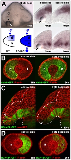
Cell movements into the neural retina can occur independent of Fgf-dependent nasal-temporal patterning. (A) Live embryo at 10ss, 3 h after Fgf8 bead implantation (arrowhead) below the ventral optic vesicle leaflet (top left). Predicted Fgf distribution along the dorsal-ventral axis of the optic vesicle in wt control and after Fgf8 bead implantation (bottom left). Ectopic foxg1 expression (top) and repression of foxd1 (bottom) in the ventral optic vesicle at 10ss, after Fgf8 bead implantation (left) compared to the control side of the same embryo (right). (B) Nasal cldnb:GFP expression (green) at 36 h on the control side (left) and the double-nasal Fgf8 bead implantation side (right) of the same embryo (red: F-actin counterstain). (C) Live images of HGn42A:GFP expression (green), colabeled with membrane-targeted RFP (red) at 20ss after Fgf8 bead implantation (left: control side, right: bead implantation side, arrowheads: distal GFP expression limit). Dotted lines: neural tube boundary. (D) Temporal HGn42A:GFP expression (green) at 36 h on the control side (left) and the double-nasal Fgf8 bead implantation side (right) of the same embryo (red: F-actin counterstain). Orientation in (A and C): cross-sections, dorsal to the top and lateral to the left; in (B and D): lateral with nasal/anterior to the left and dorsal to the top. d, dorsal optic vesicle leaflet; n, nasal; t, temporal; v, ventral optic vesicle leaflet. |