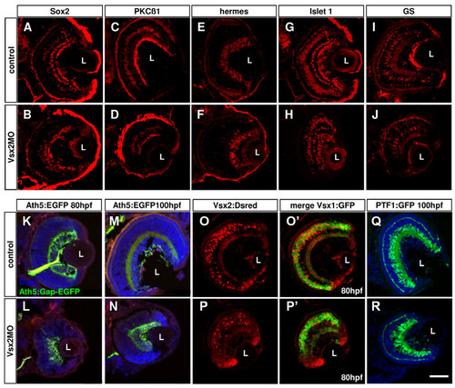
Loss of Vsx2 allows various cell types to develop in the retina. (A-J) Cryosections of 80 hours post-fertilization (hpf) retinas labelled with different cell-specific markers show that Vsx2 morphant retinas have all cell types present: Sox2 labels amacrine and Müller cells (A, B); protein kinase C (PKC)β1 labels some bipolar cells (C, D); Hermes labels ganglion cells (E, F); Islet 1 labels horizontal, bipolar, some amacrine and ganglion cells (G, H); glutamine synthetase (GS) labels Müller cells (I, J). (K-T)Cryostat sections of 80 hpf transgenic retinas similarly reveal presence of all marked cell types in Vsx2 morphants. (K-N) Vsx2 morphant Tg(ath5:Gap-GFP) retinas (double labelled with the ganglion cell marker Zn5 in red) still have green fluorescent protein (GFP)- and Zn5-labelled ganglion cells and form an optic nerve to the tectum. (O-R) Vsx2 morphant double transgenic Tg(vsx1:GFP-vsx2:dsRed) have both GFP- and DsRed-labelled cells, but appear to have comparatively fewer Vsx2:DsRed cells. As in the control transgenics, Vsx1:GFP and Vsx2:DsRed do not co-localise in the same cells. (S, T) Vsx2 morphant Tg(ptf1a:GFP) retinas show GFP labelling for horizontal and amacrine similar to that in controls, although some GFP cells are displaced and can be found closer to the lens in the GCL of the morphant retinas. L, lens. Scale bar: (A-T) 47 μm.
|
