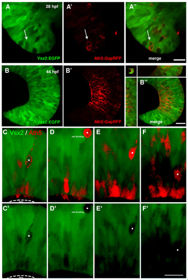
Ath5 progenitor cells arise from Vsx2 progenitors that lose Vsx2:GFP expression. (A, B) Images of live double transgenic Tg(vsx2:GFP/ath5:GapRFP) whole-mount retinas at different time points of retina development indicated as hours post-fertilisation (hpf). (A) At 28 hpf, a few cells in the retina no longer express green fluorescent protein (GFP) driven by vsx2. Some (white arrows), but not all of these Vsx2:GFP-negative cells express Ath5:RFP. (A′-B′) As development progresses, the number of cells that express Ath5:RFP increase as the number of Vsx2:GFP cells diminishes. Ath5-expressing, Vsx2:GFP-negative cells divide apically and many of the differentiating cells can be seen to settle in the developing ganglion cell layer, which becomes devoid of Vsx2:GFP cells. Images are from the movie in Additional data file 5. (C-F) Double label from a Tg(ath5:RFP;vsx2:GFP) retina in which an Ath5:RFP progenitor divides once to produce one daughter that differentiates as an RGC. (E′-F′) Vsx2 expression can be seen to be downregulated in this Ath5:RFP progenitor (whose soma is marked by the white dot). Scale bars: (A) 16 μm; (B) 27 μm; (C-F) 20 μm.
|
