
Fig. 8
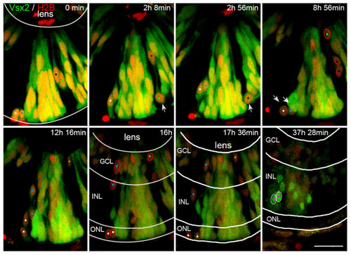
Some Vsx2 progenitors lose Vsx2 expression and differentiate as presumed photoreceptors, amacrine cells and ganglion cells. Time-lapse images of Additional file 4, showing transplanted cells from a transgenic Tg(vsx2:GFP) embryo that was injected with H2B:RFP RNA to mark all cells transplanted. Initially, all transplanted progenitor cells express Vsx2. Some cells (white arrows) undergo cell division and downregulate Vsx2:GFP expression (marked by white arrows at mitosis and white, blue, green and black spots). The Vsx2-negative daughter cells of these divisions (red outlines) end up in the ONL (presumed photoreceptors), inner INL (presumed amacrine cells) and GCL (presumed ganglion cells). In contrast, cells that upregulate the Vsx2:GFP expression late during development become restricted to the INL only, where they differentiate into Vsx2-positive bipolar or Müller cells (purple dots, white outlines). GCL, ganglion cell layer; INL, inner nuclear layer; ONL, outer nuclear layer. Scale bar: 20 μm. |