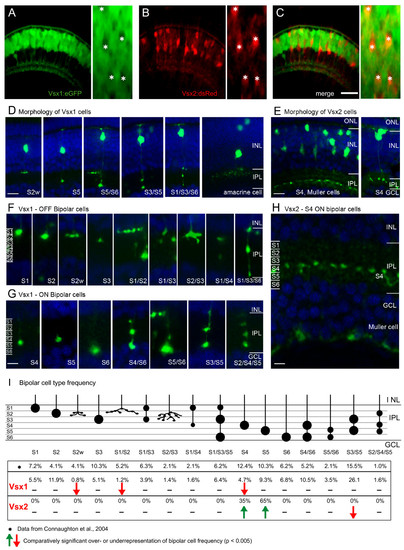
Vsx1 and Vsx2 are expressed in the distinct subtypes of bipolar cells. (A-C) Red and green fluorescence images from Tg(vsx1:GFP;vsx2:dsRed) double transgenic embryos, showing that they are expressed in separate populations of cells in the outer INL. Vsx2:dsRed positive neurons are Vsx1:GFP negative (asterisks) (D-H)Confocal images showing the morphology of individual Vsx1:GFP (D, F, G) and Vsx2:GFP (E, H) cells. Counterstained with DAPI (blue). Morphology of entire Vsx1 (D) and Vsx2 (E) expressing cells reveals bipolar cells are labelled. (F-G) Higher magnification of the IPL reveals different subtypes identified by their axon terminal stratification depth and pattern. Vsx1:GFP is expressed in many types of OFF bipolar (F) and ON bipolar (G) subtypes, which stratify throughout the entire IPL. In contrast, Vsx2:GFP is expressed in only one type of bipolar cell stratifying in stratum 4 (E, H) and in Müller cells, whose endfeet can be seen in the GCL. (I)Schematic showing the axon terminals and frequency of occurrence of Vsx1:GFP and Vsx2:GFP bipolar subtypes together with the previously described frequency from diOlistic labelling [29]. Three types of Vsx1:GFP cells occur at a statistically lower frequency than described previously and may be underrepresented in the Vsx1:GFP-labelled population (red arrows). The two types of Vsx2:GFP cells (S4 and S5), occur at a statistically higher frequency in the Vsx2:GFP population than the diOlystic population (green arrows). GCL, ganglion cell layer; INL, inner nuclear layer; IPL, inner plexiform layer; ONL, outer nuclear layer. For quantification of subtypes, IPL was subdivided into six equal strata (S1–S6). Scale bars: (A-C) 40 μm; (D, E) 10 μm; (F-H) 5 μm.
|
