
Fig. 7
- ID
- ZDB-FIG-080331-17
- Publication
- Volkmann et al., 2008 - The zebrafish cerebellar rhombic lip is spatially patterned in producing granule cell populations of different functional compartments
- Other Figures
- All Figure Page
- Back to All Figure Page
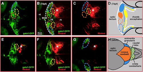
Rhodamine-dextran retrograde labeling of crista cerebellaris projections discriminates between granule cells of the vestibulo- and non-vestibulocerebellar system. Rhodamine-dextran was microinjected into one half of the GFP-fluorescent crista cerebellaris (white asterisk) in transgenic gata1:GFP embryos at 6 dpf. (A–C) Maximum intensity projections of 3-D image stack (148 μm, 75 images at 2 μm distance) recorded by laser scanning confocal microscopy at 7 dpf reveal many rhodamine-dextran labeled cells in areas of the ventrolateral (blue dashed circle) and dorsoposterior (yellow dashed circle) gata1:GFP granule cell clusters. (E–G) Single optical sections indeed revealed co-labeling of GFP-cells with rhodamine-dextran in these granule cell populations. In contrast, rhodamine-dextran labeled cells were never found in the area or to co-localize with cells of the dorsomedial granule cell cluster (orange dashed circle). (D, H) Schematic drawing of granule cell projections deduced from rhodamine-dextran retrograde labeling; note that granule cells of the dorsomedial cluster (marked orange) do not project into the crista. These studies identify the embryonic ventrolateral (D, blue dashed circle) and dorsoposterior (D, yellow dashed circle) gata1:GFP granule cell clusters as granule cells of the adult vestibulocerebellar system in zebrafish (H, schematic drawing of dorsal view) formed by the eminentia granularis (blue) and the caudal lobe (yellow), respectively. Abbr.: cb, cerebellum; MHB, midbrain–hindbrain boundary; ot, optic tectum; rh, rhombencephalon. |
| Gene: | |
|---|---|
| Fish: | |
| Anatomical Terms: | |
| Stage: | Days 7-13 |
Reprinted from Developmental Biology, 313(1), Volkmann, K., Rieger, S., Babaryka, A., and Köster, R.W., The zebrafish cerebellar rhombic lip is spatially patterned in producing granule cell populations of different functional compartments, 167-180, Copyright (2008) with permission from Elsevier. Full text @ Dev. Biol.