Fig. 6
- ID
- ZDB-FIG-080331-16
- Publication
- Volkmann et al., 2008 - The zebrafish cerebellar rhombic lip is spatially patterned in producing granule cell populations of different functional compartments
- Other Figures
- All Figure Page
- Back to All Figure Page
|
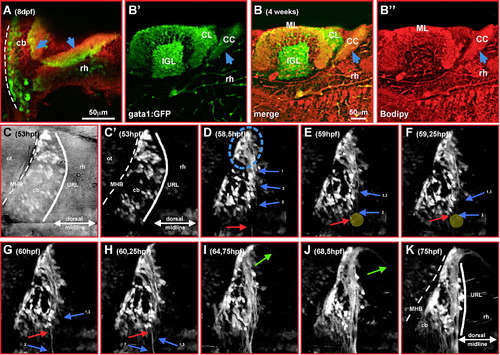
gata1:GFP granule cells project commissural axons into the crista cerebellaris. (A) Optical section of the cerebellum of 8 dpf gata1:GFP transgenic larvae. Note the dorsoposterior projection out of the cerebellum (blue arrow). Sagittal sectioning of 4-week-old gata1:GFP cerebella, immunohistochemistry against GFP-expression (B′) and counterstaining with Bodipy 630/650-X (B″) reveal positioning of these projections within the crista cerebellaris (B–B″, blue arrow). (C–K) 3-D time-lapse analysis (dorsal view) to characterize axonal projections into the crista cerebellaris (60 μm stacks of 21 individual images spaced 3 μm were recorded every 12 min). Brightest point projections of images of individual time-points are displayed. Individual axons are marked with arrows; note the contact and avoidance of growing axons from opposite cerebellar halves close to the dorsal midline (E, F, yellow circle; see slow motion sequence at the end of Movie 1). Around 65 hpf first projections into the forming crista cerebellaris can be observed (see green arrows in Movie 1 in supplementary material). Abbr.: cb, cerebellum; CC, crista cerebellaris; CL, caudal lobe; IGL, internal granule cell layer; MHB, midbrain–hindbrain boundary; ML, molecular layer; ot, optic tectum; rh, rhombencephalon; URL, upper rhombic lip. |
| Gene: | |
|---|---|
| Fish: | |
| Anatomical Terms: | |
| Stage Range: | Long-pec to Days 21-29 |
Reprinted from Developmental Biology, 313(1), Volkmann, K., Rieger, S., Babaryka, A., and Köster, R.W., The zebrafish cerebellar rhombic lip is spatially patterned in producing granule cell populations of different functional compartments, 167-180, Copyright (2008) with permission from Elsevier. Full text @ Dev. Biol.

