
Fig. 1
- ID
- ZDB-FIG-080331-11
- Publication
- Volkmann et al., 2008 - The zebrafish cerebellar rhombic lip is spatially patterned in producing granule cell populations of different functional compartments
- Other Figures
- All Figure Page
- Back to All Figure Page
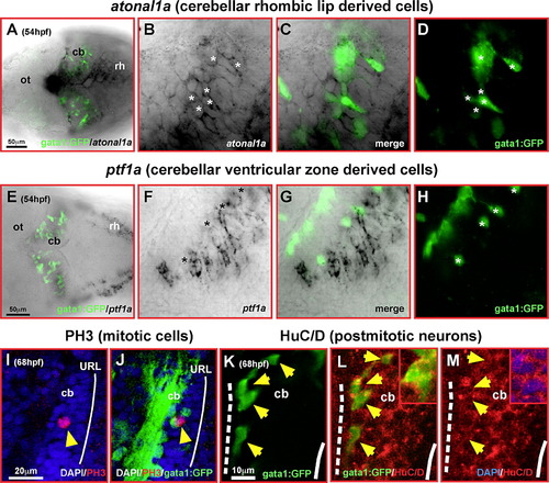
Differentiating GFP-expressing cells in the cerebellum of transgenic gata1:GFP embryos are derived from the rhombic lip. Dorsal views of cerebella (A, E) or single optical section taken by confocal microscopy of one magnified cerebellar half (B–D, F–H) analyzed by immunohistochemistry against GFP-expression and mRNA in situ hybridization for zebrafish atonal1a (A–D) or ptf1a-expression (E–H), respectively. Whereas GFP-expressing cells (D, H, white asterisk) are co-expressing atonal1a (B), they are spared by ptf1a-expression and are positioned in gaps of the ptf1a-expression pattern (F). (I–M) Single optical section of the lateral view of the cerebellum recorded by confocal microscopy at 68 hpf. Although only few GFP-expressing cells (I, J, yellow arrowhead) co-express the mitotic M-phase marker PH3 close to the upper rhombic lip (white solid line), most GFP-expressing neuronal precursors (K–M, yellow arrowheads) co-express the neuronal postmitotic marker HuC/D (L, see also inset) close to the MHB (white dashed line; note that GFP is localized throughout th28white solid line), most GFP-expressing neuronal precursors (K–M, yellow arrowheads) co-express the neuronal postmitotic marker HuC/D e cell, while HuC is confined to the cytoplasm as shown by the DAPI nuclear counterstain in the inset of panel M). Abbr.: cb, cerebellum; MHB, midbrain–hindbrain boundary; ot, optic tectum; rh, rhombencephalon; URL, upper rhombic lip. |
| Genes: | |
|---|---|
| Fish: | |
| Anatomical Terms: | |
| Stage Range: | Long-pec to Pec-fin |
Reprinted from Developmental Biology, 313(1), Volkmann, K., Rieger, S., Babaryka, A., and Köster, R.W., The zebrafish cerebellar rhombic lip is spatially patterned in producing granule cell populations of different functional compartments, 167-180, Copyright (2008) with permission from Elsevier. Full text @ Dev. Biol.