
Fig. 4
- ID
- ZDB-FIG-080331-14
- Publication
- Volkmann et al., 2008 - The zebrafish cerebellar rhombic lip is spatially patterned in producing granule cell populations of different functional compartments
- Other Figures
- All Figure Page
- Back to All Figure Page
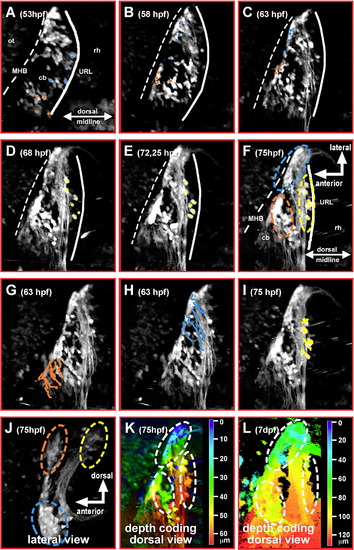
Tracing of migrating GFP-expressing granule precursor cells in gata1:GFP transgenic embryos reveals a spatial pattern of the cerebellar rhombic lip. (A–F) Dorsal view of one cerebellar half; maximum brightness projections from individual time-points of a time-lapse microscopy study of a transgenic gata1:GFP embryo are shown. Manually traced granule precursor cells contributing to the dorsomedial cluster have been marked by an orange dot, cells contributing to the ventrolateral cluster are marked by a blue dot (A–C). A smaller stationary population of GFP-expressing granule precursor cells (yellow dots) remains at the dorsoposterior edge of the differentiating cerebellum (D–F). (G–I) Independently, migratory routes were visualized by ImageJ supported cell tracing. Routes have been overlaid onto pictures of individual time-points (G, H, 63 hpf; I, 75 hpf) at which tracing has been finished. (J) Lateral view of the cerebellum, maximum intensity projection (stack of 21 images each 3 μm apart) showing that the medial and lateral granule cell clusters marked in dorsal view projections (F) are positioned apart from one another along the dorsoventrrebellum, maximum intensity projection (stack of 21 images each 3 μm apart) showing that the medial and lateral granule cell clal axis of the cerebellum. While granule precursor cells migrating towards the medial cluster (orange) remain in dorsal positions, migration of granule precursors cells heading towards the lateral cluster involves a strong ventral component. Depth coding of granule cell positions indicates that the medial and lateral granule cell clusters are about 30 to 50 μm apart along the dorsoventral axis of the cerebellum at 3 dpf (K), increasing to values of 50 to 100 μm at 7 dpf (L) due to continued growth of the cerebellum. Abbr.: cb, cerebellum; MHB, midbrain–hindbrain boundary; ot, optic tectum; rh, rhombencephalon; URL, upper rhombic lip. |
| Gene: | |
|---|---|
| Fish: | |
| Anatomical Terms: | |
| Stage Range: | Long-pec to Days 7-13 |
Reprinted from Developmental Biology, 313(1), Volkmann, K., Rieger, S., Babaryka, A., and Köster, R.W., The zebrafish cerebellar rhombic lip is spatially patterned in producing granule cell populations of different functional compartments, 167-180, Copyright (2008) with permission from Elsevier. Full text @ Dev. Biol.