
FIGURE
Fig. 7
Fig. 7
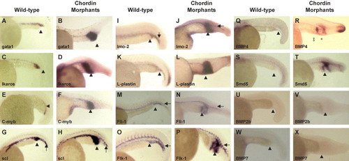
Whole-mount in-situ hybridization of WT and ChdMO embryos at 24 hpf. Expression of both hematopoietic and vascular genes, as well as BMP4 and smad5, were up-regulated in the expanded ICM (arrowhead). In the ventral tail region (arrow), there was no difference in the expression of genes between WT and the morphants. Note that BMP4 was ectopically expressed in the morphants at the tip of the yolk extension (‡) as well as the distal border of the expanded ICM (*). |
Expression Data
| Genes: | |
|---|---|
| Fish: | |
| Knockdown Reagent: | |
| Anatomical Terms: | |
| Stage: | Prim-5 |
Expression Detail
Antibody Labeling
Phenotype Data
Phenotype Detail
Acknowledgments
This image is the copyrighted work of the attributed author or publisher, and
ZFIN has permission only to display this image to its users.
Additional permissions should be obtained from the applicable author or publisher of the image.
Reprinted from Developmental Biology, 277(1), Leung, A.Y., Mendenhall, E.M., Kwan, T.T., Liang, R., Eckfeldt, C., Chen, E., Hammerschmidt, M., Grindley, S., Ekker, S.C., and Verfaillie, C.M., Characterization of expanded intermediate cell mass in zebrafish chordin morphant embryos, 235-254, Copyright (2005) with permission from Elsevier. Full text @ Dev. Biol.