
FIGURE
Fig. 8
Fig. 8
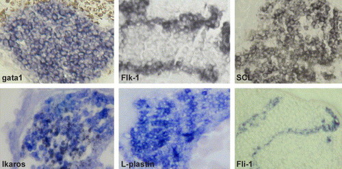
Paraffin sections (7 µm) of ChdMO embryos (24 hpf) at ICM after hybridization. Gata1, ikaros, l-plastin, and scl were generally expressed among the cells in the ICM. Vascular genes including flk1 and fli1 were expressed at the periphery of the ICM. |
Expression Data
Expression Detail
Antibody Labeling
Phenotype Data
Phenotype Detail
Acknowledgments
This image is the copyrighted work of the attributed author or publisher, and
ZFIN has permission only to display this image to its users.
Additional permissions should be obtained from the applicable author or publisher of the image.
Reprinted from Developmental Biology, 277(1), Leung, A.Y., Mendenhall, E.M., Kwan, T.T., Liang, R., Eckfeldt, C., Chen, E., Hammerschmidt, M., Grindley, S., Ekker, S.C., and Verfaillie, C.M., Characterization of expanded intermediate cell mass in zebrafish chordin morphant embryos, 235-254, Copyright (2005) with permission from Elsevier. Full text @ Dev. Biol.