
FIGURE
Fig. 4
Fig. 4
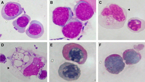
Wright staining of cytospin preparation of cells dissected out from the expanded ICM of the ChdMO embryos at 48 hpf. The ICM comprised heterogeneous population of cells, including erythroid cells (A), blast-looking cells (B), monocyte-looking cells (arrowhead, C and D). Some of the erythroid cells (E) as well as blast-looking cells (F) were PAS (Periodic Acid Schiff) staining positive. |
Expression Data
Expression Detail
Antibody Labeling
Phenotype Data
Phenotype Detail
Acknowledgments
This image is the copyrighted work of the attributed author or publisher, and
ZFIN has permission only to display this image to its users.
Additional permissions should be obtained from the applicable author or publisher of the image.
Reprinted from Developmental Biology, 277(1), Leung, A.Y., Mendenhall, E.M., Kwan, T.T., Liang, R., Eckfeldt, C., Chen, E., Hammerschmidt, M., Grindley, S., Ekker, S.C., and Verfaillie, C.M., Characterization of expanded intermediate cell mass in zebrafish chordin morphant embryos, 235-254, Copyright (2005) with permission from Elsevier. Full text @ Dev. Biol.