FIGURE SUMMARY
Title

CLN3 deficiency leads to neurological and metabolic perturbations during early development

Authors
Heins-Marroquin, U., Singh, R.R., Perathoner, S., Gavotto, F., Merino Ruiz, C., Patraskaki, M., Gomez-Giro, G., Kleine Borgmann, F., Meyer, M., Carpentier, A., Warmoes, M.O., Jäger, C., Mittelbronn, M., Schwamborn, J.C., Cordero-Maldonado, M.L., Crawford, A.D., Schymanski, E.L., Linster, C.L.
Source
Full text @ Life Sci Alliance

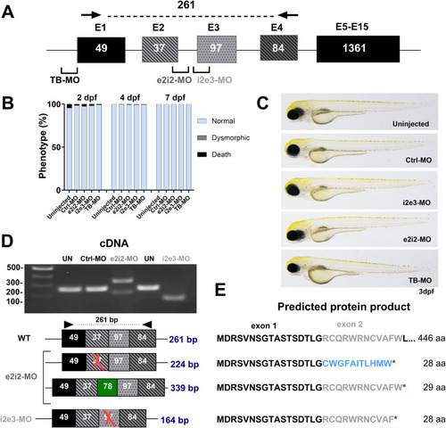
Transient knockdown of cln3 in zebrafish larvae.

(A) Schematic illustration of the cln3 gene showing the target sites for the translation-blocking (TB) and the two splice-blocking (SB; i2e3-MO and e2i2-MO) morpholinos (MOs) used in this study. Black arrows show the binding sites for the PCR primers used for SB-MO validation. (B, C) Microinjection of 8 ng SB- or TB-blocking MOs did not lead to any obvious morphological phenotype, and larvae displayed a normal development. The number of larvae analyzed at the indicated developmental time points: uninjected, 177; Ctrl-MO, 317; e2i2-MO, 250; i2e3-MO, 147; and TB-MO, 130. (D) Validation of MO efficiency by PCR amplification of target regions in cDNA of the cln3 morphants in comparison with uninjected (UN) and control morpholino–injected larvae. (E) Schematic representation of transcribed sequences and resulting translation products after treatment with the SB-blocking morpholinos. Microinjection of i2e3-MO resulted in exon 3 skipping, leading to an early stop codon. In contrast, e2i2-MO treatment led to two different splicing events, both also resulting in a premature stop codon. Excluded exons are marked with a red cross, included intron in green, and new amino acid sequence in light blue.

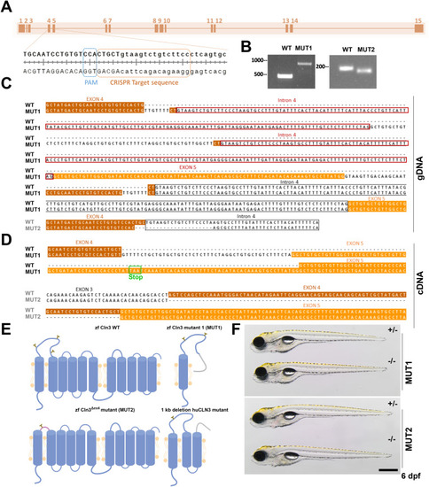
Generation of two stable cln3 mutant lines in zebrafish using CRISPR/Cas9.

(A) Schematic map of the cln3 gene with zoom on the gRNA target site. Uppercase letters represent the end of exon 4, and lowercase letters, the beginning of intron 4. (B) Gel electrophoresis of PCR amplicons from the mutated region in gDNA of F2 founders. (C) gDNA sequences of the two stable cln3 mutant lines generated by the CRISPR/Cas9 technology. (D) Based on cDNA sequence analysis from the pGEMT cloning, MUT1 carries an indel mutation resulting in a truncated translation product of 122 amino acids and MUT2 displays an in-frame deletion of exon 4. WT exon 4 sequence is highlighted in brown, exon 5 in orange, and intron 4 with a black box. Additional copies of intron 4 are highlighted with red boxes and the premature stop codon with a green box. (E) Schematic representation, based on the in silico prediction of transmembrane helices and N-glycosylation sites, of the expected protein products encoded by the WT, MUT1, and MUT2 alleles, as well as the main transcript of the most common human Batten disease CLN3 allele (1-kb deletion; huCLN3). (F) MUT1 and MUT2 mutants and heterozygous controls at 5 dpf. The scale bar represents 500 μm. Homozygous mutants are morphologically indistinguishable from their heterozygous counterparts. WT, wild type; zf, zebrafish; hu, human.

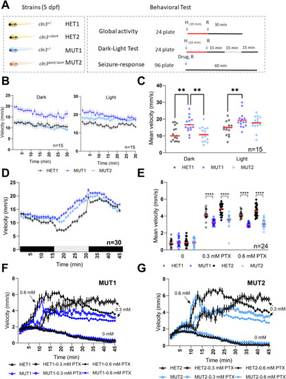
Locomotor behavior of cln3 mutant larvae under different light conditions.

(A) Experimental design of behavioral response studies for 5 dpf larvae. For the global activity and dark–light response assays, 8 larvae/line were placed in a 24-well plate (1 larva/well) and pre-adapted for 15 min to the indicated condition (light/dark) before starting the recording. The movement was tracked for 45 min in 15-min light/dark intervals or for 30 min under one continuous lightning condition. For the seizure response assay, 12 larvae/line were placed in a 96-well plate and recording was started shortly after addition of PTX for 1 h. Vehicle control was 0.6% DMSO. (B) Mean velocity (1-min intervals) of the larvae recorded under constant lighting conditions (n = 15 for each line). (B, C) Total mean velocity recorded in panel (B). (D) Mean velocity (1-min intervals) of the larvae in alternating lighting conditions (n = 30 for each line). (E) Total mean velocity in 1-h exposure to 0, 0.3, and 0.6 mM PTX. (E, F, G) Behavioral profiles (1-min intervals) of the larvae recorded in panel (E) (n = 24 for each line). All experiments shown are representative of at least three independent experiments performed on different days. In all the behavioral profiles shown, data points are means ± SEMs. In the scatter dot plots, each dot represents the total mean velocity of one larva and the red line represents the median. Statistically significant differences between lines were determined using the ordinary two-way ANOVA test followed by Sidak’s multiple comparison test (**P ≤ 0.0021 and ****P ≤ 0.0001).

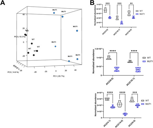
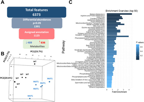
Untargeted metabolomics differentiates between WT and MUT1 samples.

(A) Experimental workflow of the untargeted metabolomics study from sample collection to data analysis. (B) MS feature filtering pipeline including statistical analyses and compound identification. Of 6,328 detected features detected in experiment 1, 1,857 differed significantly between the WT and MUT1 lines, and of those, 1,369 could be annotated. (C) Metabolite enrichment analysis of differential metabolites (P < 0.05) highlights the major dysregulated pathways between WT and MUT1 samples of experiment 1.

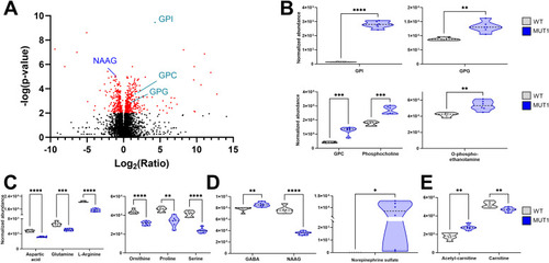
Most significantly altered metabolites between WT and MUT1 larvae based on untargeted metabolomics.

(A) Volcano plot (corrected P-value versus fold change ratio) showing significantly (P < 0.001) altered metabolites in red for experiment 1. Each dot represents one metabolite. NAAG (previously reported as a potential NCL biomarker (33)) is highlighted in blue and GPD species in turquoise. (B, C, D, E) Violin plots showing levels of selected differential metabolites as abundance values, normalized as described in the Materials and Methods section. Statistically significant differences between the WT and MUT1 lines were determined using an unpaired multiple Welch’s t test (*P ≤ 0.05; **P ≤ 0.01; ***P ≤ 0.001; and ****P < 0.0001) (n = 240 for each line, extracted and analyzed in batches of 40).

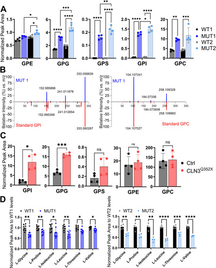
Validation of glycerophosphodiester and amino acid changes by targeted LC-MS analyses.

(A) Metabolite levels of glycerophosphodiester species (GPE, GPI, GPS, GPG, and GPC) extracted from batches of 10 whole zebrafish larvae at 5 dpf (n = 40 larvae for each genotype). (B) Representative MS/MS spectrum plots of GPI and GPC from MUT1 extracts (in blue) and metabolite standards (in red), further confirming compound identities (matching library score 83%, isotopic pattern score 100%). (A, C) Metabolite levels of the same glycerophosphodiester species as shown in panel (A), but extracted from human iPSC-derived cerebral organoids after 55 d of differentiation (n = 4). (D) Metabolite levels of amino acids differing significantly between WT and MUT1 zebrafish larvae at 5 dpf based on targeted LC-MS measurements. Data shown are means ± SDs of eight biological replicates for WT1/MUT1 (n = 200 larvae per genotype) and four biological replicates for WT2/MUT2 (n = 40 larvae per genotype). Statistically significant differences between WT and mutant zebrafish samples were determined using multiple comparison one-way ANOVA followed by Tukey’s correction, and for iPSC samples, an unpaired t test was used (*P ≤ 0.05; **P ≤ 0.01; ***P ≤ 0.001; and ns, not significant).

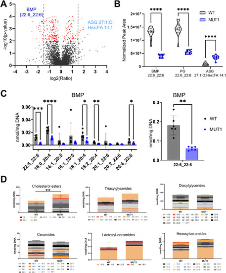
Lipidomics analysis in WT and MUT1 larvae.

(A) Volcano plot with annotated lipids found by untargeted lipidomics in non-polar extracts derived from the zebrafish larva samples generated in both experimental sets as described in the main text. Each dot represents a metabolite, and lipids with a significantly different abundance value in WT and MUT1 extracts are shown in red (P < 0.001). Gray dotted lines indicate the 1.5-fold change log2 ratio cutoff. BMP (44:12) and ASG (27:1; O;Hex;FA 14:1) are highlighted in dark and light blue, respectively. (B) Violin plots for the most significantly altered lipids based on untargeted lipidomics analysis. (C) Targeted BMP analysis in zebrafish extracts at 5 dpf. Data shown are means ± SDs for six biological replicates. Statistically significant differences between WT (gray) and MUT1 (blue) were determined using an unpaired multiple Welch’s t test (*P ≤ 0.05; **P ≤ 0.01; ***P ≤ 0.001; and ****P ≤ 0.0001). (D) Stacked bar graphs for the indicated lipid classes measured by targeted lipidomics in 5 dpf zebrafish larvae. Each stacked bar is the sum of the average amounts measured for the indicated lipid species in six biological replicates of a pool of 40 larvae, normalized against its DNA concentration. Error bars represent SDs. Statistically significant differences between both zebrafish lines were determined using a two-way ANOVA test on the summed averages for each lipid class (**P ≤ 0.01) (n = 240 larvae for each genotype).

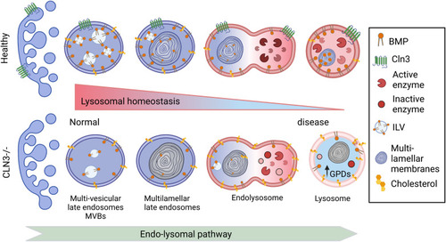
Proposed changes induced by CLN3 deficiency along the endolysosomal pathway

In healthy cells, the number of intraluminal vesicles (ILVs) increases in the early endosomes to form multilamellar bodies in the late endosomes. The latter fuse with lysosomes forming a hybrid transient endolysosome to finally form secondary lysosomes. BMP is enriched in ILVs, and its levels increase during endosome maturation, contributing to ILV formation, ceramide degradation, and cholesterol efflux (45). In CLN3-deficient cells, BMP levels are decreased, potentially leading to reduced ILV formation, as well as impaired degradation and therefore accumulation of cholesterol, glycosylceramides, and glycerophosphodiesters, all contributing to lysosomal dysfunction.

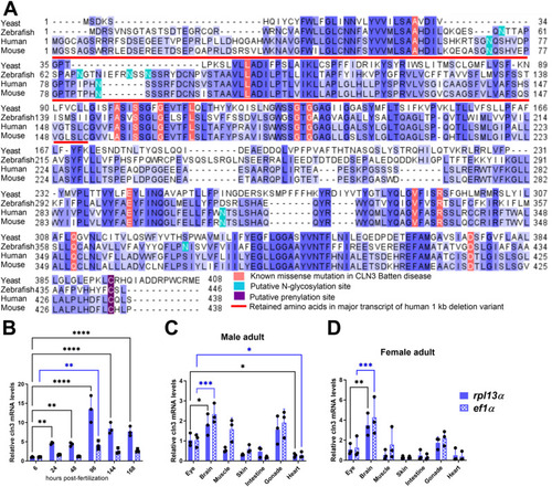
Conservation and expression of the zebrafish cln3 gene.

(A) Multiple sequence alignment of CLN3 protein sequences shows high conservation across various species (yeast, Saccharomyces cerevisiae sequence NP_012476.1; zebrafish, Danio rerio sequence NP_001007307; human, Homo sapiens sequence NP_001273038; and mouse, Mus musculus sequence NP_001139783). Gaps are indicated with dashes, and different shades of blue were used to represent the conservation level: dark blue, blue, and light blue for 100%, 75%, and 50% conservation in the selected sequences, respectively. Residues in the human sequence known to harbor missense mutations in CLN3 Batten disease are highlighted in red. The red line indicates the 153 amino acids preserved from the wild-type protein in the major transcript expressed from the 1-kb deletion allele of human CLN3. Post-translational modification sites are highlighted in green and violet. The main experimentally validated post-translational modifications are indicated (25, 26, 70). For the zebrafish Cln3 sequence, residues 1–122 may be preserved in a putative truncated expression product in the MUT1 line and residues 62–90 are missing in the predicted variant expressed by the MUT2 line described in this study. (B) Zebrafish cln3 expression levels at different developmental stages are represented relative to the expression level at 6 hpf. (C, D) Zebrafish cln3 expression levels in the indicated organs are shown relative to the expression levels in the eye. RNA was extracted from the organs of 2-yr-old male and female zebrafish for qPCR analysis. All expression levels were normalized to either the rpl13α (dark blue) or the ef1α (dotted light blue) reference genes, which were reported to be stably expressed during development and across tissues (71, 72). Data shown in panel B are technical replicates of a pool of 30 larvae. (B, C, D) Data shown are means ± SDs from three technical replicates of a pool of 30 larvae (B) or from biological replicates of three adult fish (C, D). Statistical significance was estimated using the two-way ANOVA test (*P ≤ 0.05; **P ≤ 0.01; ***P ≤ 0.001; and ****P < 0.0001). hpf, hours post-fertilization.

Generation of stable cln3 mutant lines in zebrafish using CRISPR/Cas9.

(A) Pipeline for the generation of cln3 homozygous mutants. Cas9 protein was co-injected with cln3 and slc45a2 sgRNAs into one-cell embryos. Chimeric larvae showing a pronounced albino phenotype were raised to adulthood (P0) and outcrossed with nacre fish for creating the F1 generation. F1 adult fish were screened for indel mutations in the cln3 gene. Founders with a predicted early stop codon were incrossed to create the F2 generation comprising potentially wild-type (+/+), heterozygous (+/−), and homozygous progeny (−/−). (B)cln3 sgRNA was injected into freshly fertilized embryos, and genomic DNA was extracted 24 hpf. Agarose gel analysis confirmed the high efficiency of cln3 sgRNA-Cas9 to induce indel mutations at the target site. (C) Left panel: dorsal view of a 4 dpf uninjected control wild-type and a chimeric larva. The albino phenotype can be evaluated in the eye (red arrowhead). Right panel: chimeric male (upper part) and female (lower part) P0 adults. (D) Schematic illustration of the cln3 gene showing the two primer pairs used for qPCR analysis. cln3 expression levels in MUT larvae relative to the expression levels in WT at 5 dpf. All expression levels were normalized to either the rpl13α or the ef1α reference genes. (E) Carriers were incrossed and raised to adulthood. Genotyping of F2 adult fish showed that 46% and 30% of the progeny were homozygous mutants for the nonsense and exon 4 deletion mutation, respectively. (F, G) Body length and eye area of 6 dpf larvae (n = 10 per genotype). (H) Survival curve of HET1 and MUT1 zebrafish (n = 50 per genotype).

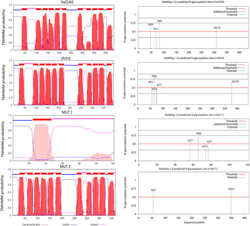
In silico prediction of transmembrane helices and N-glycosylation sites in huCLN3, zfcln3, and CRISPR mutants.

The TMHMM (v2.0) program predicts 11 putative transmembrane (helical) regions in the wild-type human and zebrafish CLN3 proteins (73Preprint). In contrast, only one transmembrane domain is predicted for the truncated protein expressed by the MUT1 line, whereas the protein encoded by the cln3Δex4 allele (MUT2 line) is predicted to have a shorter luminal loop between the first two transmembrane domains. The NetNGlyc (v1.0) tool predicts four glycosylation sites in human CLN3 (74). N49 is inside of a transmembrane domain and therefore unlikely to be glycosylated. In contrast, the zebrafish protein contains five predicted N-glycosylation sites outside of the transmembrane domains. The expression product of the MUT1 allele is predicted to conserve the first four glycosylation sites, whereas the one of the cln3Δex4 allele conserves only the first of these glycosylation sites that is placed in exon 3.

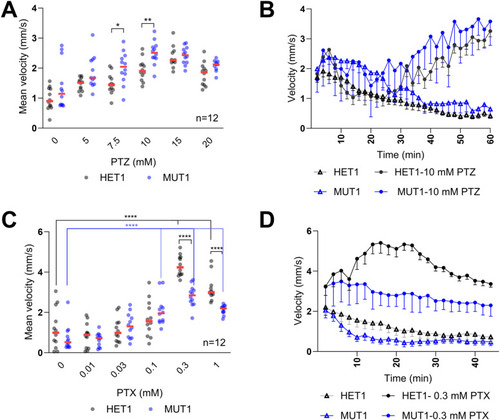
PTZ and PTX treatments in cln3-deficient zebrafish larvae.

(A) Heterozygous and homozygous mutant cln3 larvae (5 dpf) were placed in individual wells of a flat-bottom 96-well plate. Subacute (5, 7.5, and 10 mM) and acute (15 and 20 mM) concentrations of PTZ were added, and 10 min after addition of the drug (habituation), the locomotor activity was tracked and the mean velocity was calculated for each condition (40 min). (B) Average distance moved within each 2-min time bin under 0 and 10 mM PTZ yields a similar behavioral profile for both genotypes, but higher activity was observed in MUT1 larvae. Error bars represent the SEM, plotted only below the means for the sake of clarity (n = 12). (C) Similar procedure was performed with PTX as described for PTZ. (D) Behavioral profile under PTX exposure (0.3 and 1 mM). Error bars represent SDs, plotted only below the means for the sake of clarity (n = 12 for each genotype). Statistically significant differences between lines were determined using the two-way ANOVA test followed by Dunnett’s multiple comparison test (****P ≤ 0.0001).

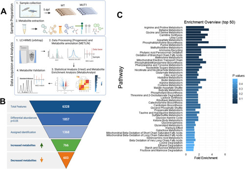
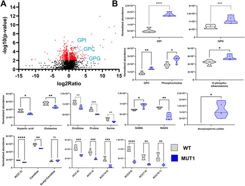
Untargeted metabolomics analysis using LC-MS (experiment 1).

(A) Principal component analysis for WT (black) and MUT1 (blue) zebrafish larvae based on all variables (m/z features) detected in positive and negative modes. The first principal component (PC1) explains 25.7% of the variation, the second component 14.9%, and the third component 10.3%. (B) Violin plots showing normalized abundance values for the indicated acylcarnitines in WT and MUT1 samples. Statistically significant differences between lines were determined using an unpaired multiple Welch’s t test (*P ≤ 0.05; **P ≤ 0.01; ***P ≤ 0.001; and ****P < 0.0001).

Multivariate and metabolite enrichment analyses of the untargeted metabolomics experiment 2.

(A) Diagram of feature filtering by statistical analysis and compound identification steps. Of 6,373 detected features, 1,125 differed significantly between the WT and MUT1 lines and could be annotated. (B) PCA using all m/z features from the polar extracts detected in positive and negative modes. The first PCA component (PC1) already shows segregation of the metabolites from WT and MUT1 samples. Each dot represents a single biological replicate of WT larvae (black) and MUT1 larvae (blue). (C) Metabolite enrichment analysis of differential metabolites (P < 0.05) highlights the major dysregulated pathways between WT and MUT1 samples in experiment 2.

Most significantly altered metabolite classes between wild-type and MUT1 larvae (experiment 2).

(A) Volcano plot (P-value versus fold change ratio) showing significantly (P < 0.001) altered metabolites in red. Each dot represents one metabolite. GPD species are highlighted in turquoise. (B) Violin plots showing normalized abundance values for the indicated metabolite values. Statistically significant differences between the two zebrafish lines were determined using an unpaired multiple Welch’s t test (*P ≤ 0.05; **P ≤ 0.01; ***P ≤ 0.001; and ****P < 0.0001).

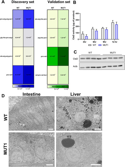
Indications for impaired proteolytic activity in MUT1 larvae.

(A) Heatmap diagrams from untargeted metabolomics experiments 1 and 2 generated from significantly different oligopeptides containing one prolyl residue. (B) CtsD activity assay in WT and MUT1 extracts showing a mild, not statistically significant, decrease in activity in the MUT1 line. (B, C) Western blot analysis of CtsD protein levels in the zebrafish extracts used for the activity assays in panel (B). (D) Representative electron micrographs of intestine and liver cells of 60-nm sagittal sections of WT and MUT1 larvae at 6 dpf. No fingerprint-like structures were observed in the MUT1 samples. Scale bars represent 3 μm.

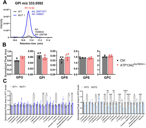
Analysis of GPDs and amino acids in polar extracts of WT, MUT1, and atp13a2sa18624−/− zebrafish larvae using targeted LC-MS analysis.

(A) Representative LC-MS extracted ion chromatograms of GPI (m/z 333,0592; RT 10.92 min) obtained with polar metabolite extracts from WT and MUT1 zebrafish larvae (5 dpf) showing the highest peak at RT 10.92 min. (B) GPD analysis in polar metabolite extracts of WT and atp13a2Sa18624−/− larvae (6 dpf). Each dot represents a pool of 10 larvae, and in total, four biological replicates were measured. (C) Amino acids that were not significantly altered in whole-larva extracts of WT and MUT, based on targeted analysis. Metabolite levels of amino acids differing significantly between WT and MUT1 zebrafish larvae at 5 dpf based on targeted LC-MS measurements. Data shown are means ± SDs of eight biological replicates for WT1/MUT1 (n = 200 larvae per genotype) and four biological replicates for WT2/MUT2 (n = 40 larvae per genotype). AA, automatic (integration) area; AH, automatic height.

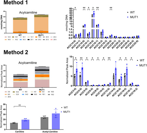
Acylcarnitine measurements using two independent targeted LC-MS methods.

In the upper part, ACs were extracted and measured by Lipometrix (Method 1). In the lower part, carnitine, acetylcarnitine, and ACs were extracted and measured at the LCSB metabolomics facility (Method 2). Each dot represents a pool of 40 larvae, and in total, six (Method 1) and four (Method 2) biological replicates were measured. In both targeted methods, AC showed slightly increased levels in MUT1 compared with WT larvae. Statistically significant differences between the zebrafish lines were determined using an unpaired parametric multiple Welch’s t test (*P ≤ 0.05; **P ≤ 0.01; ***P ≤ 0.001; and ****P < 0.0001).

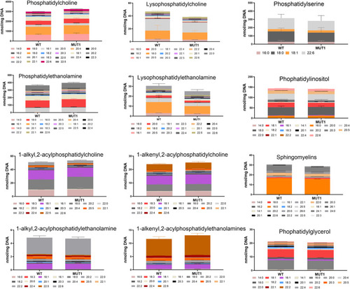
Targeted analysis of phospholipids in MUT1 larvae.

Stacked bar graphs of fatty acid composition for diverse phospholipid species assayed in non-polar extracts of 5 dpf zebrafish larvae using targeted LC-MS–based methods. Non-significant differences were observed between WT and MUT1 samples for these phospholipids. Each stacked bar is the average of six biological replicates of pools of 40 larvae, normalized against the DNA concentration.

Acknowledgments
This image is the copyrighted work of the attributed author or publisher, and ZFIN has permission only to display this image to its users. Additional permissions should be obtained from the applicable author or publisher of the image. Full text @ Life Sci Alliance