
- Title
-
A Simple, Robust, and Cost-effective Method for Genotyping Small-scale Mutations
- Authors
- Zheng, L., Hill, J., Zheng, L., Rumi, M.A.K., Zheng, X.L.
- Source
- Full text @ J Clin Transl Pathol
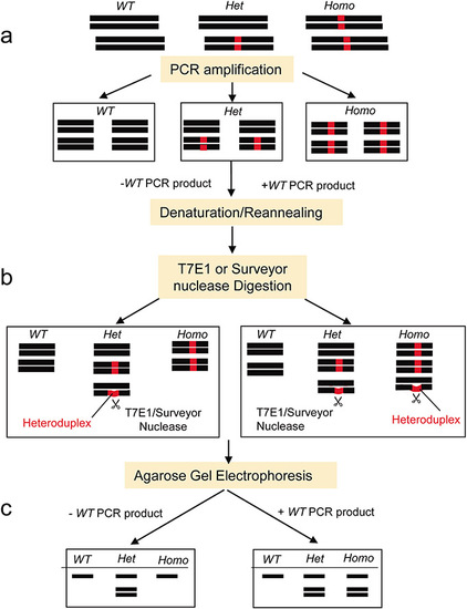
An outlined protocol for the new genotyping method.
Genomic DNA was released from the samples following tissue lysis. The cell lysate was then directly used for the PCR to amplify the target gene region. The PCR product was split into two aliquots. In one aliquot, a wild-type (WT) control PCR product was added; in another aliquot, no WT control PCR product was added. The PCR products were then denatured at 95°C and reannealed. Heteroduplexes of the DNA fragments were expected to form in the heterozygous (Het) mutant products in this step. However, when mixed with a WT control PCR product, the artificial heteroduplexes of DNA were also expected to form in the homozygous (Homo) mutant samples, but not in the wild-type samples. T7 endonuclease I (T7E1) recognized and cleaved the heteroduplexes with a mismatch ≥2 base pairs but the Surveyor nuclease could cleave a single base mismatch and indels up to at least 12 nucleotides. Thus, we could clearly differentiate the WT, Het, and Homo animals with various sizes of mutations based on the cleaved products revealed on 2% agarose gel electrophoresis. |
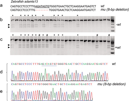
Genotyping wild-type, heterozygous, and homozygous 8 bp-deletion mutation in zebrafish adamts13.
(a) The expected wild type (wt) and mutant (mu) (8-bp deletion) sequences of zebrafish adamts13. (b–c) are the T7E1 digestion patterns of the PCR products in the absence (−wt) and presence (+wt) of the wt PCR product, respectively. Here, * and # indicate the presence of the cleavage bands (arrowheads) in the absence and presence of a wt sequence, respectively. (d–e) show the Sanger sequence tracings of wt and mu (−8 bp), respectively. |
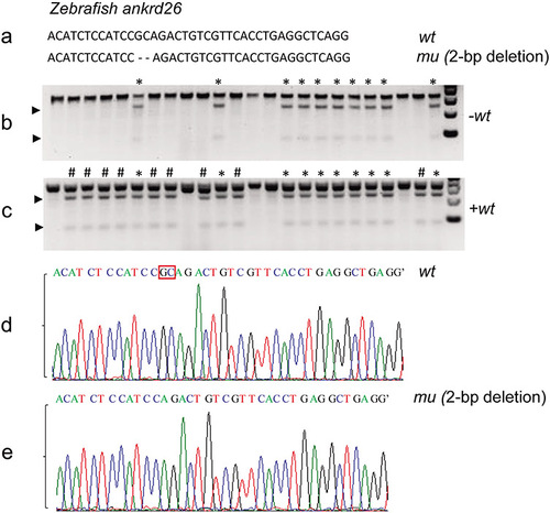
Genotyping wild-type, heterozygous, and homozygous zebrafish with a 2 bp-deletion in ankrd26.
(a) The expected DNA sequences of the wild-type (wt) and mutant (mu) (i.e., 2 bp-deletion) ankrd26. (b–c) The T7E1 digestion patterns of the PCR products in the absence and presence of a wt PCR product, respectively. Here, * and # indicate the presence of the cleavage bands (arrowheads) in the absence (−) and the presence (+) of a wt PCR sequence, respectively. (d–e) Sanger sequencing results confirmed the genotyping results (−2 bp) revealed by the agarose gel electrophoresis following T7E1 digestion. |
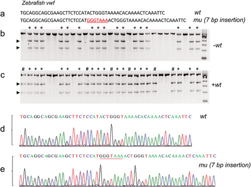
Genotyping wild-type, heterozygous and homozygous zebrafish with 7 bp-insertion in vwf.
(a) The expected DNA sequences of the wild-type (wt) and mutant (mu) (i.e., 7 bp-insertion) in zebrafish von Willebrand factor (vwf). (b–c) are the T7E1 digestion patterns of the PCR products in the absence (−) and the presence (+) of a wt PCR product, respectively. Here, * and # indicate the presence of the cleavage bands (arrowheads) in the absence and presence of a wt PCR sequence, respectively. (d–e) Sanger sequencing results confirmed the genotyping results (+7 bp-insertion) revealed by the agarose gel electrophoresis following the T7E1 digestion. |
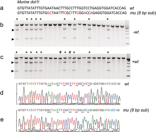
Genotyping wild-type, heterozygous, and homozygous mice with 8 bp-substitution in dot1l.
(a) The expected DNA sequences of the wild-type (wt) and mutant (mu) (i.e., 8 bp- substitution) in dot1l. (b–c) The T7E1 digestion patterns of the PCR products in the absence (−) and the presence (+) of a wt PCR product, respectively. Here, * and # indicate the presence of the cleavage bands (arrowheads) in the absence and presence of a wt PCR sequence, respectively. (d–e) Sanger sequencing of the PCR products confirmed the genotyping results (i.e., 8 bp- substitution) above. |
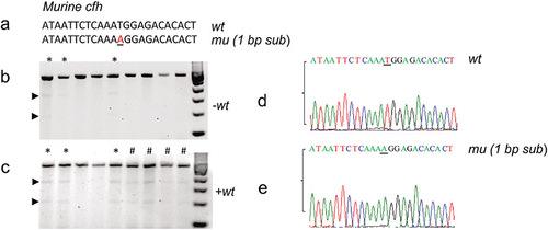
Genotyping wild-type, heterozygous, and homozygous mice with a single nucleotide substitution in cfh.
(a) The expected DNA sequences of the wild-type (wt) and mutant (mu) murine cfh. (b–c) The Surveyor nuclease digestion patterns of various PCR products in the absence (−) and the presence (+) of a wt sequence, respectively. Here, * and # indicate the presence of the cleavage bands (arrowheads) in the absence (i.e., heterozygous) and the presence (i.e., homozygous) of a wt PCR sequence, respectively. (d–e) Sanger sequencing confirmed the wt and mu (one nucleotide substitution) sequences revealed above. |