
- Title
-
Genetic compensation between Pax3 and Pax7 in zebrafish appendicular muscle formation
- Authors
- Nord, H., Kahsay, A., Dennhag, N., Pedrosa Domellöf, F., von Hofsten, J.
- Source
- Full text @ Dev. Dyn.
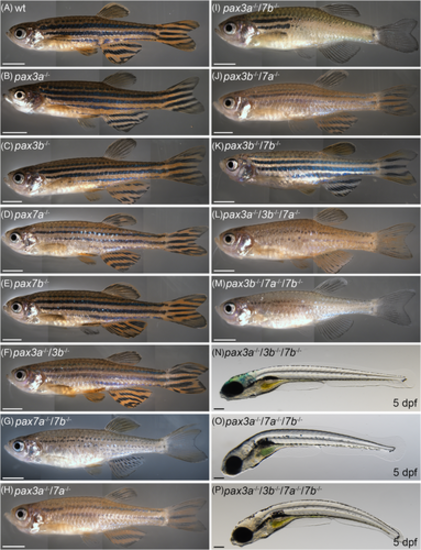
pax3 and pax7 mutant phenotypes. Lateral view of adult (3-12 months), A, wt (n = 40) and, B, pax3a−/− (n = 12), C, pax3b−/− (n = 8), D, pax7a−/− (n = 12), E, pax7b−/− (n = 17), single-mutant zebrafish and, F, pax3a−/−/3b−/− (n = 25), G, pax7a−/−/7b−/− (n = 15), H, pax3a−/−/7a−/− (n = 6), I, pax3a−/−/7b−/− (n = 6), J, pax3b−/−/7a−/− (n = 13), K, pax3b−/−/7b−/− (n = 8) double mutant zebrafish and, L, pax3a−/−/3b−/−/7a−/− (n = 4) and, M, pax3b−/−/7a−/−/7b−/− (n = 4) triple mutant zebrafish. Lateral view of, N, pax3a−/−/3b−/−/7b−/− (n = 10) and, O, pax3a−/−/7a−/−/7b−/− (n = 7) triple mutant and, P, pax3a−/−/3b−/−/7a−/−/7b−/− (n = 10) quadruple mutant zebrafish embryos at 5 dpf. Scale bar: A-M: 3 mm and N-P: 200 μm PHENOTYPE:
|
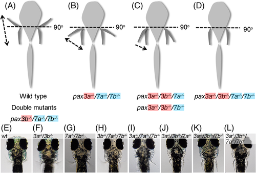
Model illustrating the fin movement of different pax3 and pax7 mutants. A, Embryos belonging to group A have the capability of moving their pectoral fins both away and toward the body, they can also move their pectoral fin in an angle >90° away from the body, group A includes wt (n = 10), all pax3/pax7 double mutant combinations (pax3a−/−/3b−/− [n = 7], pax3a−/−/7a−/− [n = 5], pax3a−/−/7b−/− [n = 5], pax3b−/−/7a−/− [n = 5], pax3b−/−/7b−/− [n = 5], and pax7a−/−/7b−/−- [n = 6]) as well as pax3b−/−/7a−/−/7b−/− triple mutant embryos (n = 11). B, Embryos in group B can move their pectoral fins both away and toward the body; however, they are unable to move their pectoral fin in an angle >90° away from the body, the triple mutant pax3a−/−/7a−/−/7b−/− (n = 5) embryos belong to group B. C, Group C represents embryos that only can move their pectoral fin toward the body and are unable to flex the fin in an angle >90° away from the body. This group comprises the triple mutant pax3a−/−/3b−/−/7a−/− (n = 10) and pax3a−/−/3b−/−/7b−/− (n = 5) embryos. D, Group D represents embryos that are unable to move their pectoral fin in any direction. This group includes the pax3a−/−/3b−/−/7a−/−/7b−/− (n = 5) quadruple zebrafish embryos. E, Snapshots representing the maximum flexed fin movement for each group is presented. pax7a and pax7b mutant alleles are highlighted in blue and pax3a and pax3b in red. Dotted line represents the 90° angle from the body axis. Arrows indicate movement of pectoral fin PHENOTYPE:
|
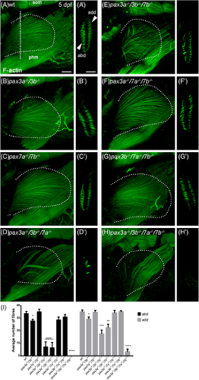
Morphology of pectoral fin muscle fibers in pax3 and pax7 mutants. Lateral view of pectoral fin of, A, wt, B, pax3a−/−/3b−/− and, C, pax7a−/−/7b−/− double mutant, D, pax3a−/−/3b−/−/7a−/−, E, pax3a−/−/3b−/−/7b−/−, F, pax3a−/−/7a−/−/7b−/− and, G, pax3b−/−/7a−/−/7b−/− triple mutant and, H, pax3a−/−/3b−/−/7a−/−/7b−/− quadruple zebrafish embryos at 5 dpf stained with phalloidin to visualize F-actin; A'-H', shows cross section of pectoral fin at the level of the vertical dotted line indicated in A. Arrowheads indicate abductor (abd) and adductor (add) muscles. The shape of the fin was determined using the 4′,6-diamidino-2-phenylindole (DAPI) channel and is outlined by a dotted line in A-H. I, Average number of muscle fibers in abductor and adductor muscle of the pectoral fin of wt (n = 7), pax3a−/−/3b−/− (n = 7), pax7a−/−/7b−/− (n = 6), pax3a−/−/3b−/−/7a−/− (n = 9), pax3a−/−/3b−/−/7b−/− (n = 5), pax3a−/−/7a−/−/7b−/− (n = 5), pax3b−/−/7a−/−/7b−/− (n = 7), and pax3a−/−/3b−/−/7a−/−/7b−/− (n = 5) zebrafish embryos at 5 dpf. Error bars indicate SEM and significance was calculated using Student's t-test where P < .05 was considered significant, *P < .05, **P < .01, ***P < .001, and ****P < .0001. abd, abductor; add, adductor; phm, posterior hypaxial muscle; som, somites. Scale bar: 50 μm PHENOTYPE:
|
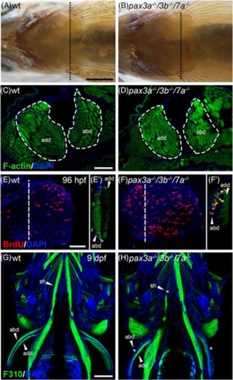
Adult pax3a−/−/3b−/−/7a−/− mutants have restored pectoral fin muscle. Ventral view of adult 12 months old, A, wt and, B, pax3a−/−/3b−/−/7a−/− mutants (n = 3) showing pectoral fin muscle. Dashed black line indicates area of cross section shown in C,D. Phalloidin staining of pectoral fin sections of 12 months old, C, wt and, D, pax3a−/−/3b−/−/7a−/− mutants. BrdU (red) incorporation between 72 and 96 hpf in pectoral fins of, E, wt (n = 51) and, F, pax3a−/−/3b−/−/7a−/− mutant (n = 7), dashed line indicates level of confocal cross sections shown in E' and F' where F-actin is visualized in green. Ventral view of 9 dpf juveniles stained with F310 antibody (green) in G wt (n = 49) and, H, pax3a−/−/3b−/−/7a−/− mutant (n = 4). Missing or reduced muscle is indicated by *. abd, abductor; add, adductor; sh, sternohyoideus. A,B, Scale bar: 1 mm, CD,G,H, 200 μm, E,F, 50 μm |
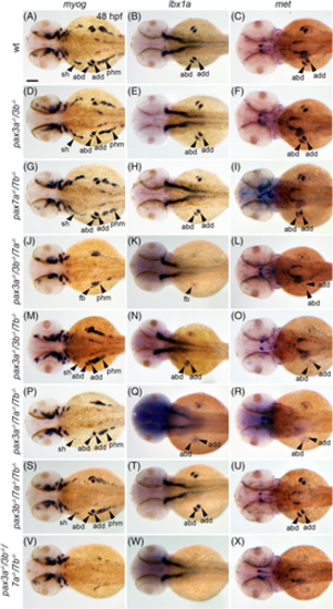
Pax3 and Pax7 are redundantly required for migrating muscle progenitor (MMP) myogenesis. Dorsal view showing the expression of myogenin (myog) (left column), ladybird homeobox 1a (lbx1a) (middle column) and met (right column) in A-C wt (n = 47), D-F, pax3a−/−/3b−/− (n = 33), G-I, pax7a−/−/7b−/− (n = 13), J-L, pax3a−/−/3b−/−/7a−/− (n = 13), M-O, pax3a−/−/3b−/−/7b−/−(n = 16), P-R, pax3a−/−/7a−/−/7b−/− (n = 12), S-U, pax3b−/−/7a−/−/7b−/− (n = 33), and V-X, pax3a−/−/3b−/−/7a−/−/7b−/− (n = 7) mutant zebrafish embryos at 48 hpf. Arrowheads indicate abductor, adductor, sternohyoideus, and posterior hypaxial muscle, or fin bud where impossible to distinguish between abductor/adductor. abd, abductor; add, adductor; fb, fin bud; sh, sternohyoideus; phm, posterior hypaxial muscle. Scale bar: 100 μm EXPRESSION / LABELING:
PHENOTYPE:
|
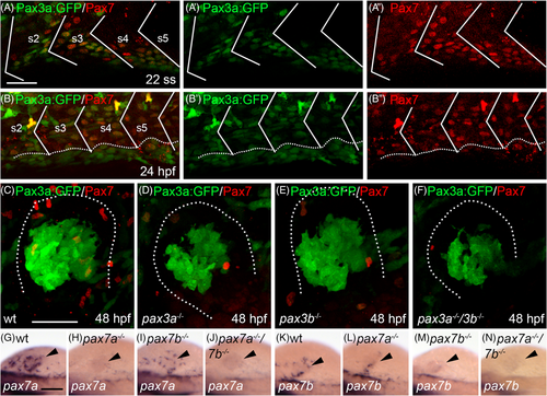
Pax7 and Pax3a:GFP are co-expressed in the somite before migrating muscle progenitors (MMPs) start to migrate out. Lateral view of somites 2-5 showing transgenic expression of A Pax3a:GFP (green) and immunohistochemical detection of Pax7 (red), A', Pax3a:GFP and, A", Pax7 in zebrafish embryos at the 22 somite stage (ss). Lateral view of somites 2-5 showing transgenic expression of B Pax3a:GFP (green) and immunohistochemical detection of Pax7 (red), B', Pax3a:GFP and, B'', Pax7 in zebrafish embryos at 24 hpf. Myosepta are marked with white lines and dashed line in B indicates yolk-somite border. Transgenic expression of Pax3a:GFP (green) and immunohistochemical detection of Pax7 (red) in C wt, D, pax3a−/−, E, pax3b−/− and, F, pax3a−/−/3b−/− double mutant pectoral fin bud at 48 hpf. Dotted line in C-F indicates outline of fin bud. Expression of pax7a in, G, wt (n = 65), H, pax7a−/− (n = 8), I, pax7b−/− (n = 18) and, J, pax7a−/−/7b−/− (n = 4). Expression of pax7b in (K) wt (n = 101), L, pax7a−/− (n = 13), M, pax7b−/− (n = 12), and N pax7a−/−/7b−/− (n = 5). Arrowhead indicates location of fin bud. Scale bar: A-F, 50 μm, G-N, 100 μm EXPRESSION / LABELING:
PHENOTYPE:
|
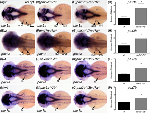
Expression of pax3 and pax7 is up-regulated in the absence of pax7 and pax3, respectively. Dorsal view showing the expression of pax3a in A wt (n = 18), B, pax7a−/−/7b−/− double mutant (n = 9) and, C pax3b−/−/7a−/−/7b−/− triple mutant embryos (n = 5) at 48 hpf. D, Relative mRNA expression levels of pax3a in wt and pax7a−/−/7b−/− double mutant embryos at 48 hpf. Dorsal view showing the expression of pax3b in E wt (n = 25), F, pax7a−/−/7b−/− double mutant (n = 8), and, G, pax3a−/−/7a−/−/7b−/− triple mutant embryos (n = 5) at 48 hpf. H, Relative mRNA expression level of pax3b in wt and pax7a−/−/7b−/− double mutant embryos at 48 hpf. Dorsal view showing the expression of pax7a in I wt (n = 23), J, pax3a−/−/3b−/− double mutant (n = 11) and, K, pax3a−/−/3b−/−/7b−/− triple mutant embryos (n = 8) at 48 hpf. L, Relative mRNA expression level of pax7a in wt and pax3a−/−/3b−/− double mutant embryos at 48 hpf. Dorsal view showing the expression of pax7b in M wt (n = 42), N, pax3a−/−/3b−/− double mutant (n = 8) and, O, pax3a−/−/3b−/−/7a−/− triple mutant embryos (n = 10) at 48 hpf. P, Relative mRNA expression level of pax7b in wt and pax3a−/−/3b−/− double mutant embryos at 48 hpf. Student's t-test was used to calculate significance (P < .05). Error bars indicate SEM. Arrowheads indicate abductor, adductor of fin bud. Abbreviations: abd, abductor; add, adductor; fb, fin bud. Scale bar: 100 μm |
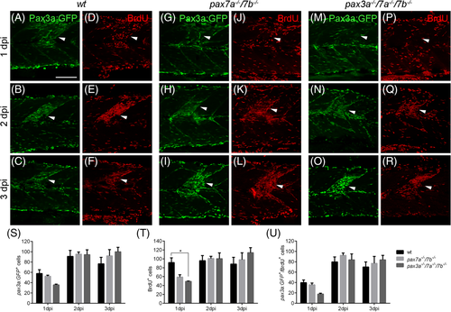
Initiation of muscle regeneration is not affected by the absence of pax7 and pax3. Transgenic expression of Pax3a:GFP (green) and incorporation of BrdU (red) in A-F wt (n = 16), G-L, pax7a−/−/7b−/− double mutant (n = 15) and, M-R, pax3a−/−/7a−/−/7b−/− triple mutant (n = 13) zebrafish embryos injured at somite number 10 at 4 dpf shown at A,D,G,J,M,P 1 dpi, B,E,H,K,N,Q, 2 dpi, and, C,F,I,L,O,R, 3 dpi. Average number of Pax3a:GFP+, BrdU+ and Pax3:GFP/BrdU double positive cells per somite at 1, 2, and 3 dpi is presented in S-U. Error bars indicate SEM and significance was calculated using Student's t-test where P < .05 was considered significant. Arrowheads indicate area of injury. Scale bar: 100 μm |