|
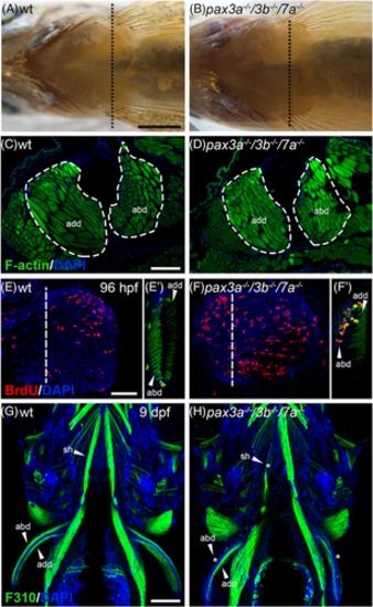
Adult pax3a−/−/3b−/−/7a−/− mutants have restored pectoral fin muscle. Ventral view of adult 12 months old, A, wt and, B, pax3a−/−/3b−/−/7a−/− mutants (n = 3) showing pectoral fin muscle. Dashed black line indicates area of cross section shown in C,D. Phalloidin staining of pectoral fin sections of 12 months old, C, wt and, D, pax3a−/−/3b−/−/7a−/− mutants. BrdU (red) incorporation between 72 and 96 hpf in pectoral fins of, E, wt (n = 51) and, F, pax3a−/−/3b−/−/7a−/− mutant (n = 7), dashed line indicates level of confocal cross sections shown in E' and F' where F-actin is visualized in green. Ventral view of 9 dpf juveniles stained with F310 antibody (green) in G wt (n = 49) and, H, pax3a−/−/3b−/−/7a−/− mutant (n = 4). Missing or reduced muscle is indicated by *. abd, abductor; add, adductor; sh, sternohyoideus. A,B, Scale bar: 1 mm, CD,G,H, 200 μm, E,F, 50 μm
|