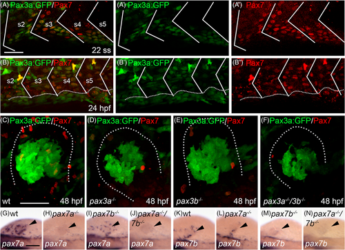
Pax7 and Pax3a:GFP are co-expressed in the somite before migrating muscle progenitors (MMPs) start to migrate out. Lateral view of somites 2-5 showing transgenic expression of A Pax3a:GFP (green) and immunohistochemical detection of Pax7 (red), A', Pax3a:GFP and, A", Pax7 in zebrafish embryos at the 22 somite stage (ss). Lateral view of somites 2-5 showing transgenic expression of B Pax3a:GFP (green) and immunohistochemical detection of Pax7 (red), B', Pax3a:GFP and, B'', Pax7 in zebrafish embryos at 24 hpf. Myosepta are marked with white lines and dashed line in B indicates yolk-somite border. Transgenic expression of Pax3a:GFP (green) and immunohistochemical detection of Pax7 (red) in C wt, D, pax3a−/−, E, pax3b−/− and, F, pax3a−/−/3b−/− double mutant pectoral fin bud at 48 hpf. Dotted line in C-F indicates outline of fin bud. Expression of pax7a in, G, wt (n = 65), H, pax7a−/− (n = 8), I, pax7b−/− (n = 18) and, J, pax7a−/−/7b−/− (n = 4). Expression of pax7b in (K) wt (n = 101), L, pax7a−/− (n = 13), M, pax7b−/− (n = 12), and N pax7a−/−/7b−/− (n = 5). Arrowhead indicates location of fin bud. Scale bar: A-F, 50 μm, G-N, 100 μm
|
