
Fig. 3
- ID
- ZDB-FIG-220127-5
- Publication
- Nord et al., 2021 - Genetic compensation between Pax3 and Pax7 in zebrafish appendicular muscle formation
- Other Figures
- All Figure Page
- Back to All Figure Page
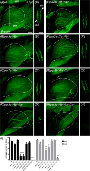
Morphology of pectoral fin muscle fibers in pax3 and pax7 mutants. Lateral view of pectoral fin of, A, wt, B, pax3a−/−/3b−/− and, C, pax7a−/−/7b−/− double mutant, D, pax3a−/−/3b−/−/7a−/−, E, pax3a−/−/3b−/−/7b−/−, F, pax3a−/−/7a−/−/7b−/− and, G, pax3b−/−/7a−/−/7b−/− triple mutant and, H, pax3a−/−/3b−/−/7a−/−/7b−/− quadruple zebrafish embryos at 5 dpf stained with phalloidin to visualize F-actin; A'-H', shows cross section of pectoral fin at the level of the vertical dotted line indicated in A. Arrowheads indicate abductor (abd) and adductor (add) muscles. The shape of the fin was determined using the 4′,6-diamidino-2-phenylindole (DAPI) channel and is outlined by a dotted line in A-H. I, Average number of muscle fibers in abductor and adductor muscle of the pectoral fin of wt (n = 7), pax3a−/−/3b−/− (n = 7), pax7a−/−/7b−/− (n = 6), pax3a−/−/3b−/−/7a−/− (n = 9), pax3a−/−/3b−/−/7b−/− (n = 5), pax3a−/−/7a−/−/7b−/− (n = 5), pax3b−/−/7a−/−/7b−/− (n = 7), and pax3a−/−/3b−/−/7a−/−/7b−/− (n = 5) zebrafish embryos at 5 dpf. Error bars indicate SEM and significance was calculated using Student's t-test where P < .05 was considered significant, *P < .05, **P < .01, ***P < .001, and ****P < .0001. abd, abductor; add, adductor; phm, posterior hypaxial muscle; som, somites. Scale bar: 50 μm |