
- Title
-
Characterization of Pax3 and Sox10 transgenic Xenopus laevis embryos as tools to study neural crest development
- Authors
- Alkobtawi, M., Ray, H., Barriga, E.H., Moreno, M., Kerney, R., Monsoro-Burq, A.H., Saint-Jeannet, J.P., Mayor, R.
- Source
- Full text @ Dev. Biol.
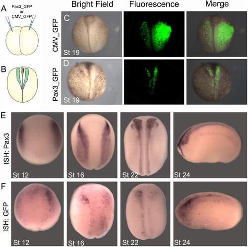
Generation of Pax3-GFP transient transgenic embryos.(A) pax3 or CMV promoters fused to GFP were injected into Xenopus embryos as described to generate transient transgenic embryos. (B) GFP fluorescence was examined at neurula stage (stage 19). (C) CMV-GFP transient transgenic embryo, showing ubiquitous expression. (D) Pax3-GFP transient transgenic embryo showing expression restricted to the neural folds. (E, F) In situ hybridization (ISH) for comparison of endogenous Pax3 expression with GFP expression in Pax3-GFP transient transgenic embryos. (E) ISH for pax3 at the indicated stages. (F) ISH for GFP in Pax3-GFP embryos at the indicated stages. Note the similar expression of Pax3 and GFP. 92% of Pax3-GFP embryos exhibited expression in the neural folds (n= 210). |
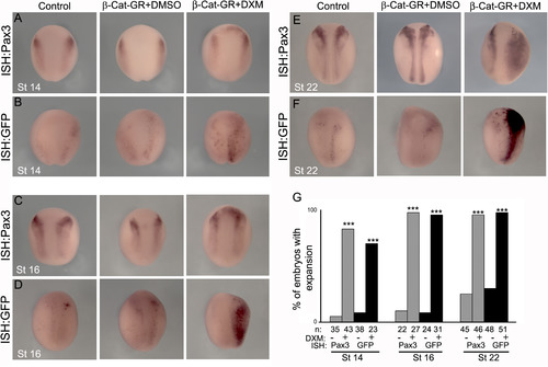
Pax3-GFP responds to Wnt signaling. (A, C, E) ISH for endogenous Pax3 in wild type embryos at the indicated stages. (B, D, F) ISH for GFP in Pax3-GFP transient transgenic embryos at the indicated stages. Embryos were non-injected (Control) or injected with an inducible form of β-catenin (β-Cat-GR) and treated with DXM (in DMSO) to induce β-catenin, or DMSO alone. (G) Quantification of the percentage of embryos that showed expansion in Pax3/GFP expression. Note that activation of β-catenin leads to an equivalent increase of Pax3 and GFP expression. *** P<0.001. |
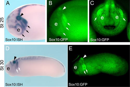
Characterization of Sox10-GFP stable transgenic embryos. (A-C) Stage 25 embryos. (A) ISH for endogenous sox10 in wild-type embryos at the indicated stage, side view. (B, C) GFP fluorescence of Sox10-GFP stable transgenic embryos at the indicated stage, B, side view and C, anterior view. (D, E) Stage 30 embryos, side view. (D) ISH for endogenous sox10 in wild type embryos at the indicated stage. (E) GFP fluorescence of Sox10-GFP stable transgenic embryos at the indicated stage. Note the similar expression of Sox10 and GFP in the migrating neural crest cells (arrows). Arrow head indicates the otic vesicle and e the eye. |
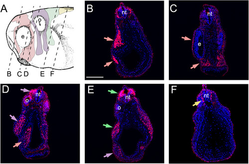
GFP expression in migrating neural crest in Sox10-GFP stable transgenic embryos. (A) Diagram indicating the level of the transverse sections shown in panels B-F. Pink shows mandibular neural crest; Purple shows hyoid neural crest; Green shows branchial neural crest; Yellow shows trunk/vagal neural crest. (B-F) Sections of Sox10-GFP stable transgenic embryos after immunostaining for GFP and DAPI. (B, C) Mandibular neural crest. (D) Hyoid and mandibular neural crest. (E) Branchial and hyoid neural crest. (F) Trunk/vagal neural crest. e: eye; o- otic vesicle; nt: neural tube. Epidermal immunostaining is likely to be non-specific background. |
Reprinted from Developmental Biology, 444 Suppl 1, Alkobtawi, M., Ray, H., Barriga, E.H., Moreno, M., Kerney, R., Monsoro-Burq, A.H., Saint-Jeannet, J.P., Mayor, R., Characterization of Pax3 and Sox10 transgenic Xenopus laevis embryos as tools to study neural crest development, S202-S208, Copyright (2018) with permission from Elsevier. Full text @ Dev. Biol.