
|
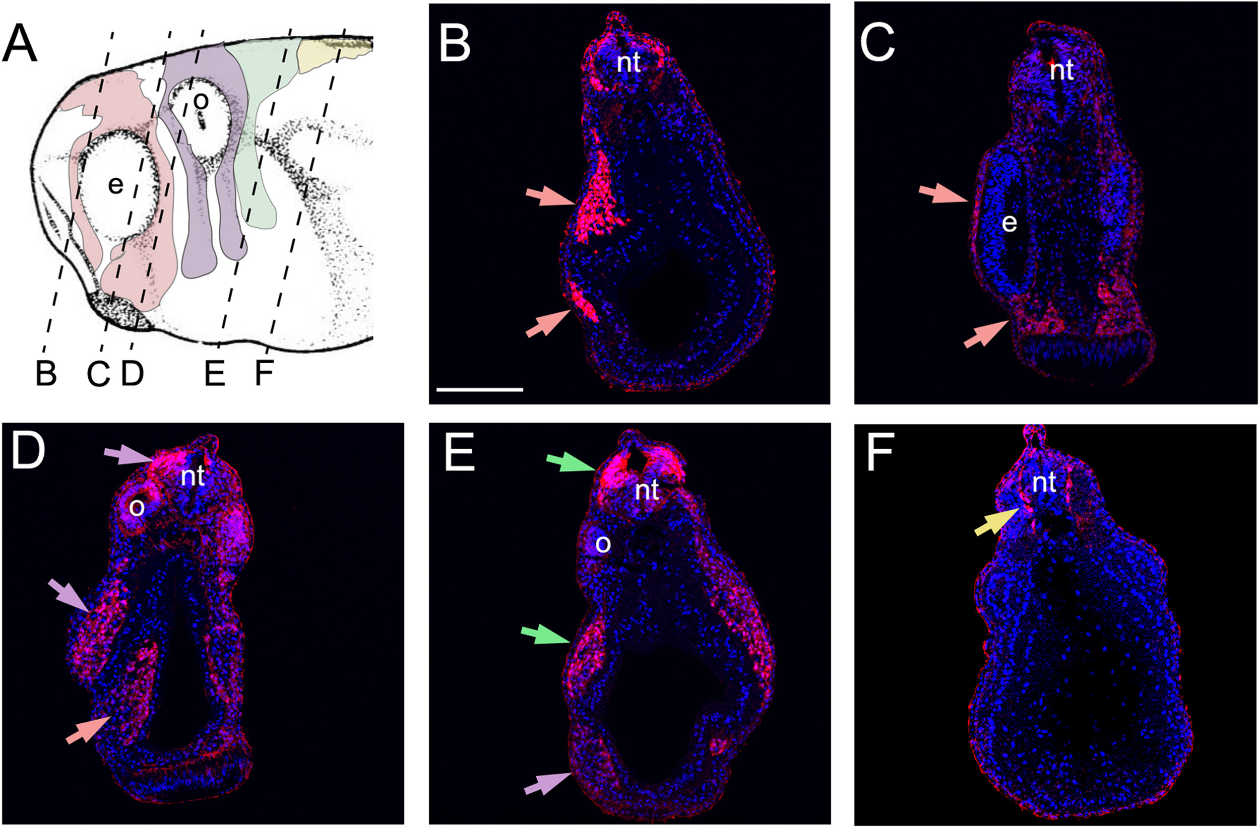
Fig. 6
GFP expression in migrating neural crest in Sox10-GFP stable transgenic embryos. (A) Diagram indicating the level of the transverse sections shown in panels B-F. Pink shows mandibular neural crest; Purple shows hyoid neural crest; Green shows branchial neural crest; Yellow shows trunk/vagal neural crest. (B-F) Sections of Sox10-GFP stable transgenic embryos after immunostaining for GFP and DAPI. (B, C) Mandibular neural crest. (D) Hyoid and mandibular neural crest. (E) Branchial and hyoid neural crest. (F) Trunk/vagal neural crest. e: eye; o- otic vesicle; nt: neural tube. Epidermal immunostaining is likely to be non-specific background.
Reprinted from Developmental Biology, 444 Suppl 1, Alkobtawi, M., Ray, H., Barriga, E.H., Moreno, M., Kerney, R., Monsoro-Burq, A.H., Saint-Jeannet, J.P., Mayor, R., Characterization of Pax3 and Sox10 transgenic Xenopus laevis embryos as tools to study neural crest development, S202-S208, Copyright (2018) with permission from Elsevier. Full text @ Dev. Biol.