
|
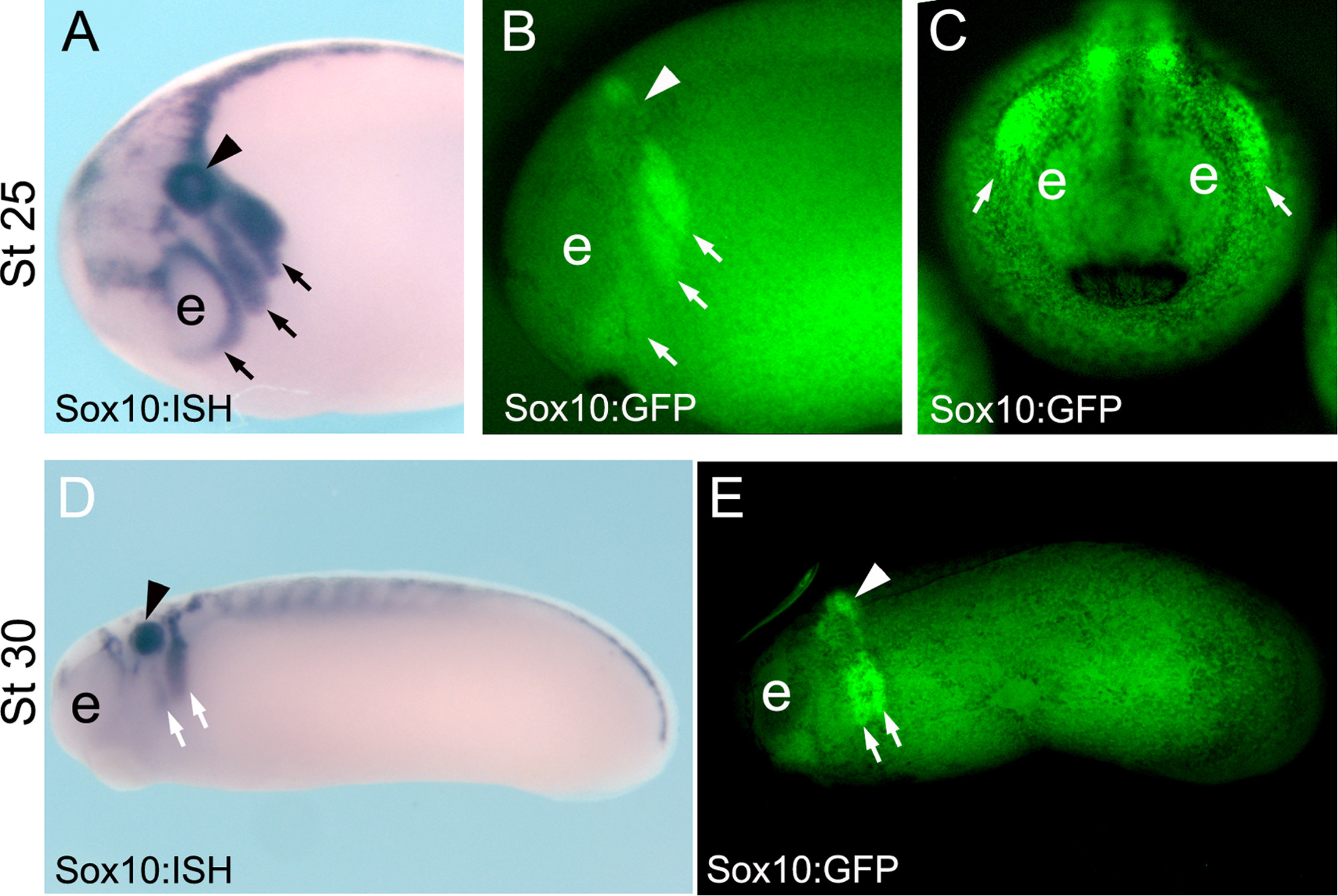
Fig. 5
Characterization of Sox10-GFP stable transgenic embryos. (A-C) Stage 25 embryos. (A) ISH for endogenous sox10 in wild-type embryos at the indicated stage, side view. (B, C) GFP fluorescence of Sox10-GFP stable transgenic embryos at the indicated stage, B, side view and C, anterior view. (D, E) Stage 30 embryos, side view. (D) ISH for endogenous sox10 in wild type embryos at the indicated stage. (E) GFP fluorescence of Sox10-GFP stable transgenic embryos at the indicated stage. Note the similar expression of Sox10 and GFP in the migrating neural crest cells (arrows). Arrow head indicates the otic vesicle and e the eye.
Reprinted from Developmental Biology, 444 Suppl 1, Alkobtawi, M., Ray, H., Barriga, E.H., Moreno, M., Kerney, R., Monsoro-Burq, A.H., Saint-Jeannet, J.P., Mayor, R., Characterization of Pax3 and Sox10 transgenic Xenopus laevis embryos as tools to study neural crest development, S202-S208, Copyright (2018) with permission from Elsevier. Full text @ Dev. Biol.