
FIGURE
Fig. 3
- ID
- ZDB-FIG-190715-6
- Publication
- Alkobtawi et al., 2018 - Characterization of Pax3 and Sox10 transgenic Xenopus laevis embryos as tools to study neural crest development
- Other Figures
- All Figure Page
- Back to All Figure Page
Fig. 3
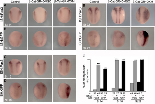
Pax3-GFP responds to Wnt signaling. (A, C, E) ISH for endogenous Pax3 in wild type embryos at the indicated stages. (B, D, F) ISH for GFP in Pax3-GFP transient transgenic embryos at the indicated stages. Embryos were non-injected (Control) or injected with an inducible form of β-catenin (β-Cat-GR) and treated with DXM (in DMSO) to induce β-catenin, or DMSO alone. (G) Quantification of the percentage of embryos that showed expansion in Pax3/GFP expression. Note that activation of β-catenin leads to an equivalent increase of Pax3 and GFP expression. *** P<0.001. |
Expression Data
Expression Detail
Antibody Labeling
Phenotype Data
Phenotype Detail
Acknowledgments
This image is the copyrighted work of the attributed author or publisher, and
ZFIN has permission only to display this image to its users.
Additional permissions should be obtained from the applicable author or publisher of the image.
Reprinted from Developmental Biology, 444 Suppl 1, Alkobtawi, M., Ray, H., Barriga, E.H., Moreno, M., Kerney, R., Monsoro-Burq, A.H., Saint-Jeannet, J.P., Mayor, R., Characterization of Pax3 and Sox10 transgenic Xenopus laevis embryos as tools to study neural crest development, S202-S208, Copyright (2018) with permission from Elsevier. Full text @ Dev. Biol.