
- Title
-
The Role of her4 in Inner Ear Development and Its Relationship with Proneural Genes and Notch Signalling
- Authors
- Radosevic, M., Fargas, L., Alsina, B.
- Source
- Full text @ PLoS One
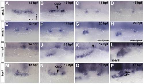
Spatiotemporal expression of proneural genes at early stages of inner ear development. (A–D) In situ hybridization in wild-type embryos for atoh1b. atoh1b is expressed in a large medial domain adjacent to the hindbrain at 12 hpf (A) to then progressively restrict to two patches that correspond to the future anterior and posterior prosensory domains (B–D). Between 13 hpf and 14 hpf atoh1b is downregulated at the central medial domain (CMD) (arrow in B). (E–H) In situ hybridization in wild-type embryos for atoh1a. atoh1a is expressed in the anterior and posterior sensory domains by 13 hpf and 14 hpf (E, F). At 20 hpf the expression in the anterior domain increases (G) and just two positive cells remain in the posterior (H). (I–K) In situ hybridization in wild-type embryos for neurog1. Expression of neurog1 in the inner ear starts at 15 hpf (J) and it is extended from an anterolateral position to a posteromedial domain (K). neurog1 progressively invades the CMD (arrow in K). (L–P) In situ hybridization in wild-type embryos for her4. her4 expression is found at 12 hpf (M) to become restricted to the future sensory maculae by 13 hpf (N). At 16 hpf her4 presents higher expression levels (O) and at 17 hpf, the expression of her4 appears in the neurogenic domain being a sum of the expressions from both domains (arrows in P). By 20 hpf the expression only remains at the anterior sensory domain (L). Dorsal views; anterior to the left, medial to the top. Dashed circles delineate otic vesicles. All images are at same magnification. Scale bar: 25 µm. EXPRESSION / LABELING:
|
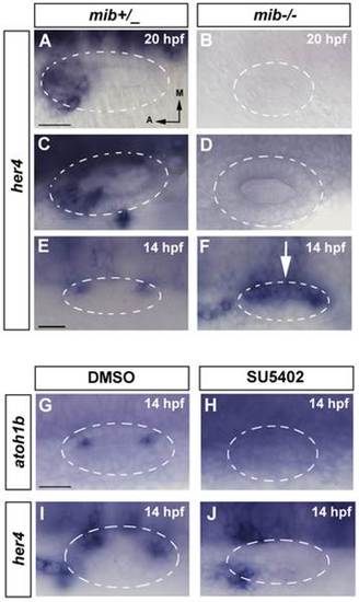
her4 is regulated by Notch in the neurogenic domain but only partially in the sensory domain. (A–F) In situ hybridization for her4 expression in control and mib-/- embryos. (A, C, E) her4 expression in wild-type embryos. At 14 hpf her4 expression is restricted to the two sensory patches (E). At 20 hpf and 24 hpf her4 expression is detected in the sensory and neurogenic domains (A, C). (B, D, F) her4 expression in mib-/- embryos. At 14 hpf her4 expression is detected in the anterior and posterior sensory domains, but it is not repressed in the CMD (white arrow in F). At 20 hpf and 24 hpf its expression in the sensory and neurogenic domains is completely abolished (B, D). (G–J) In situ hybridization for atoh1b and her4 at 14 hpf in DMSO and SU5402 treated embryos. atoh1b (G, H) and her4 expression (I–J) expression is completely lost in embryos treated with SU5402 (H,J) compared with control embryos treated with DMSO (G,I). Dorsal views; anterior to the left, medial to the top. Dashed circles delineate otic vesicles. Images A–D, E–F and G–J are at same magnification. All scale bars: 25 µm. EXPRESSION / LABELING:
|
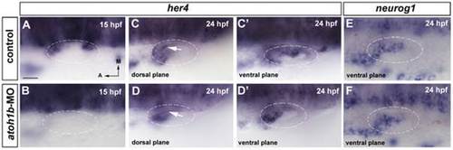
her4 expression requires atoh1b in the sensory domain. (A–D′) In situ hybridization for her4 expression in wild-type and atoh1b-MO injected embryos. (A, B) In 15 hpf control embryos, her4 expression is detected in the future anterior and posterior sensory domains (A), while in morphant embryos her4 expression is lost (B). (C, D) At 24 hpf her4 expression is also lost in the sensory territory in morphant embryos (arrow in D) compared with controls (arrow in C). (C′, D′) The expression in the neurogenic domain is not affected in morphant embryos. (E, F) In situ hybridization for neurog1 expression in wild-type (E) and atoh1b-MO injected (F) embryos. neurog1 expression in atoh1b morphant embryos is not affected. Dorsal views; anterior to the left, medial to the top. Dashed circles delineate otic vesicles. All images are at same magnification. Scale bar: 25 µm. EXPRESSION / LABELING:
|
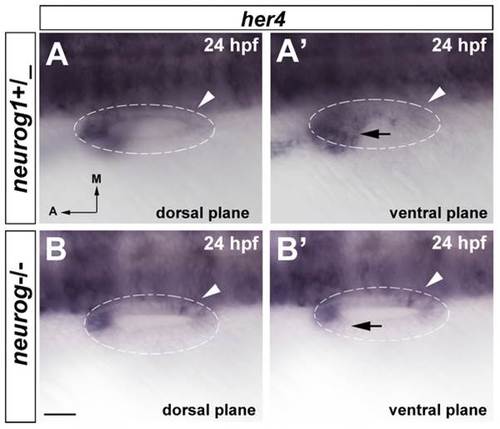
neurog1 is required to induce her4 expression in the neurogenic domain. (A, A′) In situ hybridization for her4 expression in wild-type embryos. her4 expression is detected in both, the anterior sensory domain and the neurogenic domain (arrow A′), but not in the posterior sensory domain (arrowheads A, A′). (B–B′) In situ hybridization for her4 expression in neurog1-/- embryos. Her4 is not expressed in the neurogenic domain (arrow B′) and her4 expression in the posterior sensory domain is detected (arrowheads B, B′). Dashed circles delineate otic vesicles. All images are at same magnification. Scale bar: 25 µm. |
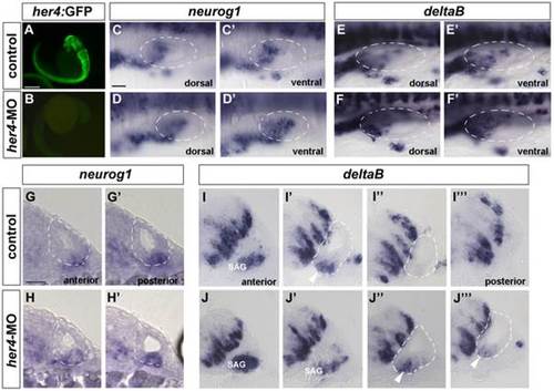
Increased density of neurog1 and deltaB-positive cells in the neurogenic domain after her4 blockade. (A, B) GFP expression in Tg(her4:GFP) line. In her4-MO injected embryos GFP expression is completely lost (B) compared to controls (A). (C–D′) Dorsal views of 24 hpf control (C, C′) and her4-MO injected (D, D′) embryos stained by in situ hybridization for neurog1. (D, D′) neurog1 expression in her4 morphant embryos is increased in the ventral plane (D′) compared to controls (C′). (E–F′) Dorsal views of 27 hpf control (E, E′) and her4-MO injected (F, F′) embryos stained by in situ hybridization for deltaB. (F, F′) deltaB expression in her4 morphant embryos is increased in the dorsal and ventral plane (F, F′) compared to controls (E, E′). (G–H′) Sequential transversal sections; medial to the left, dorsal to the top of 24 hpf control (G, G′) and her4-MO injected (H, H′) embryos stained by in situ hybridization for neurog1. In morphant embryos an increased number of cells in the otic epithelium stained for neurog1 is observed compared to controls. (I–J′′′) Sequential transversal sections; medial to the left, dorsal to the top of 27 hpf control (I–I′′′) and her4-MO injected (J, J′′′) embryos stained by in situ hybridization for deltaB. In morphant embryos the number of deltaB-positive cells in the otic epithelium (compare J′′ with I′) is increased and also the size of the SAG (J, J′). In control embryos SAG is only present in one section (I). Arrowheads point to epithelial neuroblasts. Dashed circles delineate otic vesicles. Scale bar in A and B: 500 µm, Scale bar in C-F′: 25 µm, Scale bar G-J′′′: 25 µm. |
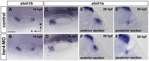
Blockade of her4 has no increase on atoh1b and atoh1a expression. (A–B) Dorsal views of 14 hpf control (A) and her4-MO injected (B) embryos stained by in situ hybridization for atoh1b. atoh1b expression is not affected in the prosensory domains in her4-MO injected embryos. (C–D) Dorsal views of 24 hpf control (C) and her4-MO injected (D) embryos stained by in situ hybridization for atoh1a. atoh1a expression decreases in her4-MO injected embryos. (E–F′) Transversal sections anterior (E, F) and posterior sections (E′, F′) of 28 hpf control (E, E′) and her4-MO injected (F, F′) embryos stained by in situ hybridization for atoh1a. atoh1a expression is as wild-type in her4-MO injected embryos. Dashed circles delineate otic vesicles. All images are at same magnification. Scale bar: 25 µm. EXPRESSION / LABELING:
|
expression is restricted to the anterior and posterior sensory domains from its onset and is not induced at the CMD. |